
|
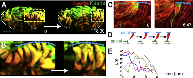
Fig. 5
Cells Shorten at Late Stages of Optic Vesicle Evagination
(A and B) Time-lapse movie starting at around 8–9 ss (z stack projection, six z slices, 5 μm intervals) of evaginating optic vesicles in an embryo mosaically labeled with Kaede.
(C) Time-lapse movie starting at around 8–9 ss of a cell expressing the fusion protein Pard3-GFP in the eye field of an embryo ubiquitously expressing a membrane localized RFP fusion protein, as it shortens (green arrows).
(D) Schematic of the labeled cell in (C).
(E) Quantification of cell length over time (five cells were followed from around 6–7 ss in two embryos; cell length was measured at around every 5–6 min).
White (A) and blue (B and C) dotted lines outline the edge of the optic vesicles, red dotted lines in (A) highlight the midline of the embryo, and yellow dotted boxes in (A) correspond to the regions shown in (B). Scale bars, 64 μm (A) and 27 μm (C).
Reprinted from Developmental Cell, 27(3), Ivanovitch, K., Cavodeassi, F., and Wilson, S.W., Precocious acquisition of neuroepithelial character in the eye field underlies the onset of eye morphogenesis, 293-305, Copyright (2013) with permission from Elsevier. Full text @ Dev. Cell