
|
Fig. 2
Two Morphologically Distinct Cell Populations within the Forming Optic Vesicles
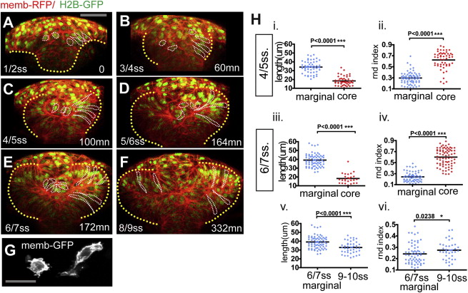
(A–F) Time-lapse movie from neural plate-1 ss (t = 0 min, 10.5 hpf) up to 8–9 ss (t = 332 min, around 13.5 hpf) of an embryo ubiquitously expressing membrane-RFP (red) and H2B-GFP (green). The yellow dotted line highlights the neural anlage at t = 0 (A) and the eye field/optic vesicles at subsequent time points. Example marginal and core cells are outlined in white (B–F). Detailed morphology is not evident at this magnification but can be seen in (G) and in other figures. Scale bar, 64 μm (A).
(G) Membrane-GFP labeling of typical core and marginal cells at 4 ss. Scale bar, 27 μm.
(H) Quantification of the cell length (i, iii, and v) and the cell rdn index (ii, iv, and vi) of marginal (blue) and core (red) cells. Black bars indicate mean (Mann-Whitney U test). Numbers of cells quantified are as follows: (i) cell length at 4–5 ss, n = 56 marginal cells in eight embryos and n = 48 core cells in five embryos; (ii) rdn index at 4–5 ss, n = 71 marginal cells in eight embryos and n = 62 core cells in five embryos; (iii) cell length at 6–7 ss, n = 73 marginal cells in eight embryos and n = 26 core cells in five embryos; (iv) rdn index at 6–7 ss, n = 63 marginal cells in eight embryos and n = 53 core cells in five embryos; (v) marginal cell length at 9–10 ss, n = 58 cells in five embryos; and (vi) marginal cell rdn at 9–10 ss, n = 38 cells in four embryos.
In order to distinguish unambiguously the shape of single eye field cells, in three-dimensions, large z stacks (60 μm in average) were acquired on mosaically labeled whole-mount embryos. See also Movie S2.
Reprinted from Developmental Cell, 27(3), Ivanovitch, K., Cavodeassi, F., and Wilson, S.W., Precocious acquisition of neuroepithelial character in the eye field underlies the onset of eye morphogenesis, 293-305, Copyright (2013) with permission from Elsevier. Full text @ Dev. Cell