
IMAGE
Fig. 6
- ID
- ZDB-IMAGE-140327-7
- Publication
- Lee et al., 2003 - The zebrafish forkhead transcription factor Foxi1 specifies epibranchial placode-derived sensory neurons
- All Figures
- Figures for Lee et al., 2003
Image
|
Figure Caption
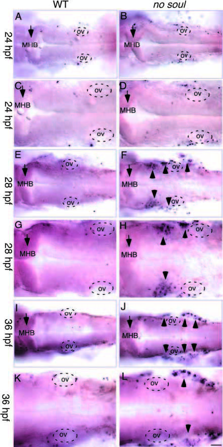
Fig. 6 TUNEL staining of whole-mount wild-type (left) and no soul mutant (right) embryos. C,D,G,H,K,L are higher magnification views of A,B,E,F,I,J, respectively. (A-D) 24 hpf embryos showing comparable TUNEL staining in wild-type and no soul mutant embryos. (E-H) 28 hpf embryos showing increased TUNEL staining in the anterior lateral cranial placodal region. (I-L) 36 hpf embryos showing increased TUNEL staining in the posterior cranial placodal region. MHB, mid-hindbrain boundary; ov, otic vesicle. Scale bar: 100 μm (A,B,E,F,I,J), 50 μm (C,D,G,H,K,L).
Figure Data
Acknowledgments
This image is the copyrighted work of the attributed author or publisher, and
ZFIN has permission only to display this image to its users.
Additional permissions should be obtained from the applicable author or publisher of the image.
Full text @ Development