
|
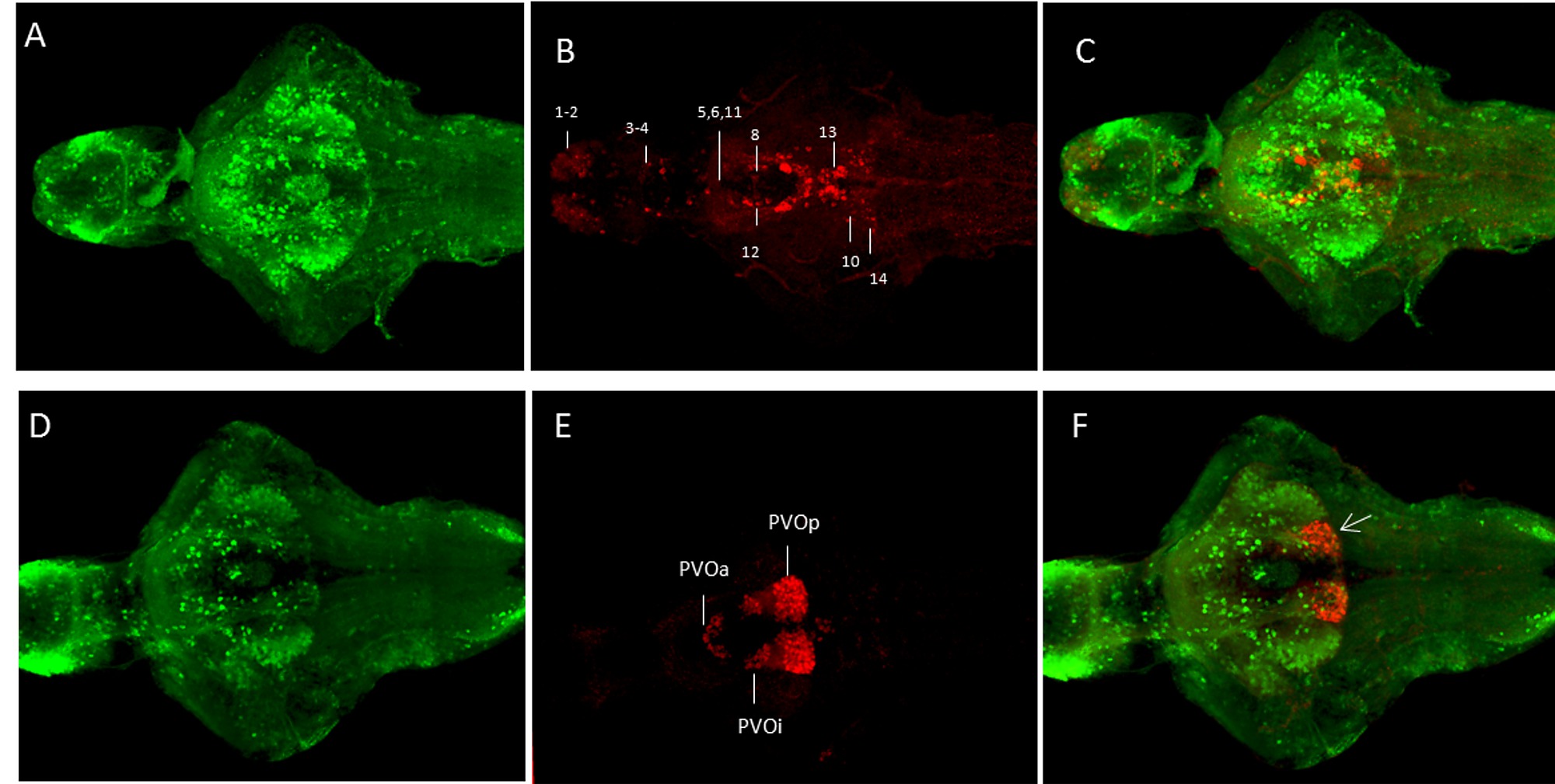
Fig. S1 Comparison of the GFP expression pattern with TH-ir and 5-HT-ir in Tg(pink1:EGFP) fish at 7 dpf. D-F. TH ir compared to GFP distribution in the Tg(pink1:EGFP) fish. GFP is more widely distributed than TH-ir. A ventral view of the maximum projection images for TH and GFP. The cell populations and numbering are as in [28].
G-I. The 5-HT-ir is colocalized with GFP expression in the posterior recess of the paraventricular organ (arrowhead).
5-HT – 5-hydroxy tryptophan, TH – tyrosine hydroxylase, ir – immunoreactivity, GFP – Green fluorescent protein, PVOa – paraventricular organ anterior part, PVOi – paraventricular organ intermediate part, PVOp – paraventricular organ posterior part.
Scale bar represents 100 μm.