
Fig. S1
- ID
- ZDB-IMAGE-140324-13
- Publication
- Samson et al., 2013 - 3-OST-7 regulates BMP-dependent cardiac contraction
- All Figures
- Figures for Samson et al., 2013
|
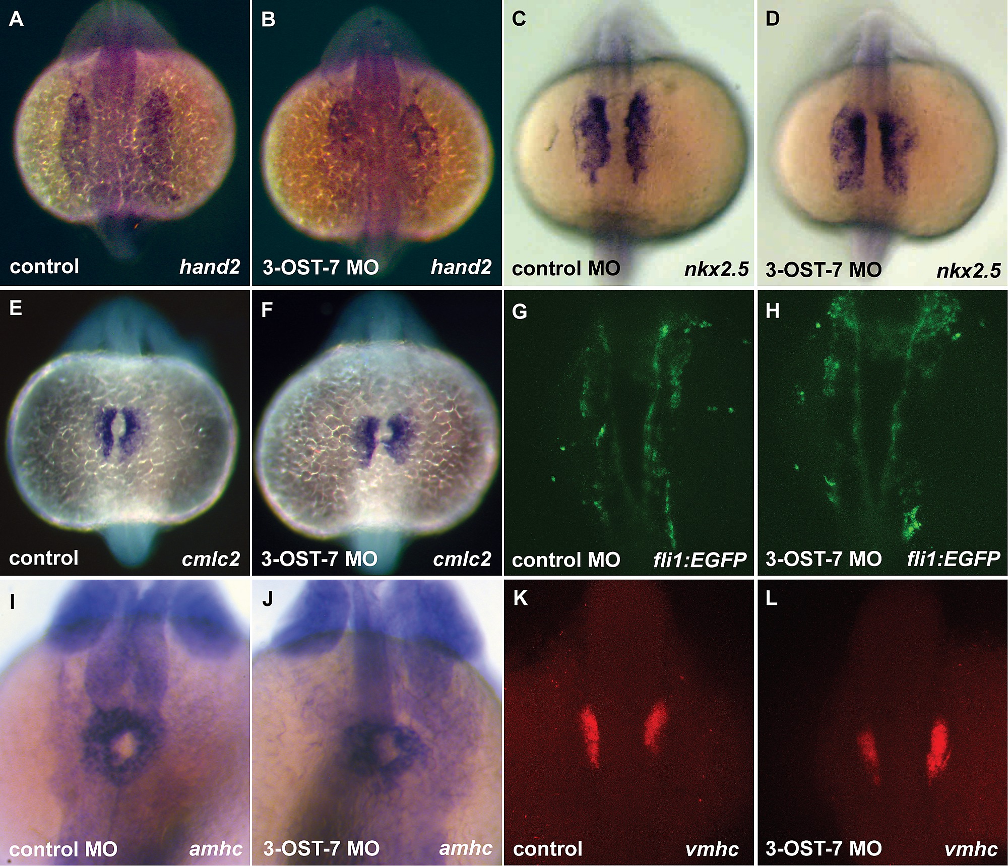
Fig. S1
Heart field specification proceeds normally in 3-OST-7 morphants. Dorsal views (anterior on top) of control (uninjected, wild-type) (A, C, E, and K), control MO (injected with control 3-OST-3Z MO) (G and I), and 3-OST-7 morphant (B, D, F, H, J and L) embryos; n = 35 for each group. ISHs for: lateral plate mesoderm marker hand2 (A and B, 17 hpf) and cardiac precursor cell marker nkx2.5 (C and D, 17 hpf), myocardial precursor cell marker cmlc2 (E and F, 17 hpf), atrial precursor cell marker amhc (I and J, 20 hpf), and ventricular precursor cell marker vmhc (K and L, 18 hpf) showed comparable levels and patterns of expression in control and 3-OST-7 morphant embryos. Imaging of fli1 expression in Tg(fli1:EGFP) zebrafish at 18 hpf revealed endocardial lineage is intact in 3-OST-7 morphant embryos (G and H).