
Fig. 3
- ID
- ZDB-IMAGE-140320-32
- Publication
- Link et al., 2000 - The zebrafish young mutation acts non-cell-autonomously to uncouple differentiation from specification for all retinal cells
- All Figures
- Figures for Link et al., 2000
|
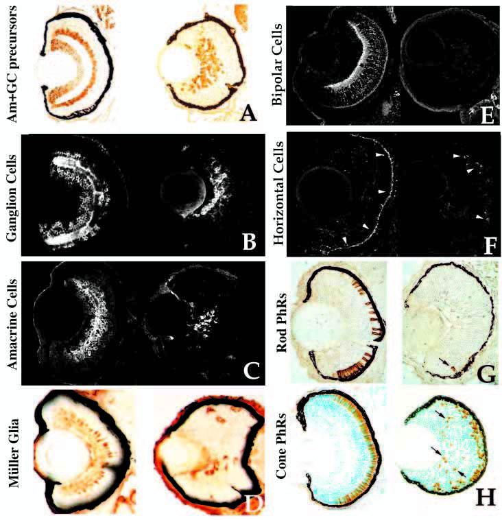
Fig. 3 Retinal cell type marker analysis in young mutants. Central, transverse retinal sections of wild-type (left) and young mutant (right) siblings assessed for celltype- specific markers (see Text and Table 1 for details). (A) Hu antigen (ganglion and amacrine cells and their precursors), 3 dpf. (B) 7A11 antigen (ganglion cells and their axons), 3 dpf. (C) 5E11 antigen (amacrine cells and their processes), 3 dpf. (D) Carbonic anhydrase II immunoreactivity (Muller glial cells), 4 dpf. Carbonic anhydrase II is also expressed in non-ocular ectoderm. (E) Protein kinase C immunoreactivity (bipolar cells and processes), 4 dpf. (F) HCII. 7 immunoreactivity (arrowheads) (horizontal cells and their processes), 3.5 dpf. (G) ID1 antigen (rod photoreceptors), 4 dpf. Arrow indicates rod cell in the ventral patch of a mutant. (H) Zn-1 antigen (red and green cone photoreceptors], 4 dpf. Arrows indicate ectopic, mispatterned photoreceptors in the young retina.