Fig. 2
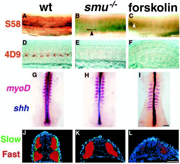
Fig. 2 smu-/- and forskolin-treated embryos have defects in slow muscle fiber type development. (A-C) Side views of S58 antibody labeled 24h embryos. S58 specifically labels slow muscle fibers. In wild-type embryos (A) approximately 20 slow muscle fibers span each chevron-shaped somite very clearly. In smu-/- (B) and forskolin-treated (C) embryos, slow muscle is markedly reduced; individual slow muscle fibers are marked with arrowheads. (D-F) Side views of 4D9 Engrailed antibody labeling of muscle pioneer slow muscle fibers at 24h. 4D9 labels 2-5 muscle pioneer nuclei per somite in wild-type (D), while smu-/- (E) and forskolin-treated (F) embryos have a complete loss of muscle pioneer slow muscle cells. (G-I) Dorsal view of 12h-14h embryos hybridized to show myoD (red) and shh (dark blue) expression. MyoD is expressed in wildtype embryos (G) in the somites and in the adaxial cells of the presomitic mesoderm. smu-/- (H) and forskolintreated (I) embryos express myoD in the somites, but do not have any expression in cells adjacent to the notochord. (J-L) Transverse sections through the trunk of 30h embryos labeled with S58 (green) and zm4 (red) monoclonal antibodies to identify slow and fast muscle fibers, respectively. In sections of wild-type embryos (J), slow muscle fibers form a monolayer around the deeper fast muscle fibers. smu-/- (K) and forskolin-treated (L) embryos have a nearly complete loss of S58 labeling throughout the entire trunk, whereas zm4 staining is still present. Bars, 50 μm (A-F); 50 μm (G-I); 100 μm (J-L).

