
Fig. 3
- ID
- ZDB-IMAGE-140319-12
- Publication
- Zeller et al., 1999 - The zebrafish diwanka gene controls an early step of motor growth cone migration
- All Figures
- Figures for Zeller et al., 1999
|
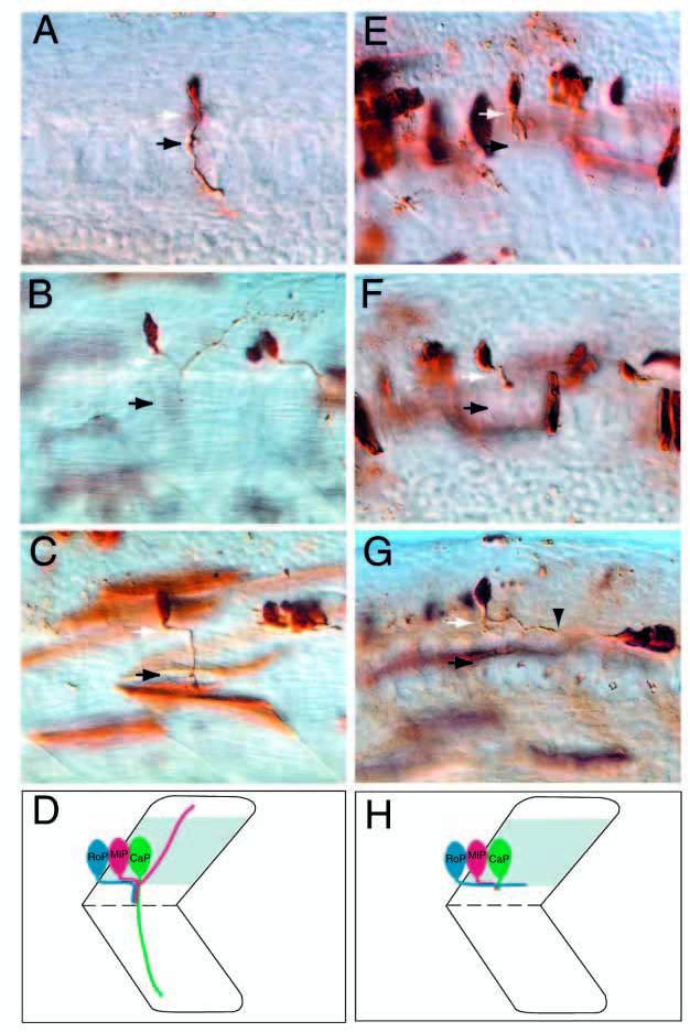
Fig. 3 diwanka is required for motor axon migration from the spinal cord to the somites. Stereotypic axonal projections of CaP (A), MiP (B) and RoP (C) neurons in wild-type embryos. White arrows point to the lower end of the spinal cord (out of focal plane) and black arrows point to the choice point. In diwanka mutants, all CaP axons (E) and MiP axons (F) extend normally within the spinal cord, but are severely affected as they migrate on the common path (black arrow). (G) All RoP axons in diwanka mutants complete the longitudinal path within the spinal cord, but most fail to exit the spinal cord and instead project further caudally within the spinal cord (arrowhead points to the growth cone). Note that the longitudinal projection of the mutant RoP axon is about twice the length of the corresponding projection of wild-type RoP in Fig. 4C.