
|
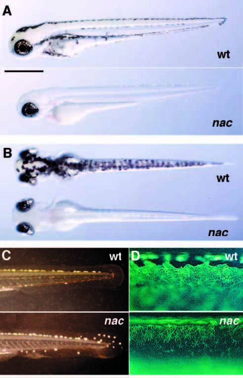
Fig. 1 The nacre mutant. Lateral (A) and dorsal (B) views of wildtype (top) and nacre (bottom) larvae at 3 days postfertilization. nacre homozygotes are missing all neural-crest-derived melanophores, but pigmentation of the eye is normal. (C) Tail iridophores, viewed with epi-illumination. nacre mutants (bottom) have increased numbers of iridophores, including many in the tail fin. (D) Xanthophore pigmentation, viewed under UV light, is slightly reduced in nacre mutants (bottom). All visible fluorescence is due to xanthophores, while the dark patches in the top panel are melanophores. Scale bar is approximately 500 μm in A-C, 100 μm in D.