
Fig. 3
- ID
- ZDB-IMAGE-140317-4
- Publication
- Takke et al., 1999 - her1, a zebrafish pair-rule like gene, acts downstream of notch signalling to control somite development
- All Figures
- Figures for Takke et al., 1999
|
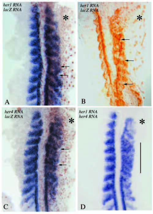
Fig. 3 Somitic defects following injection of her1 (A,B), her4 (C) or her1 and her4 (D) RNA. (A,C,D) In situ hybridizations with MyoD; (B) an anti-myosin staining. A-C have also been processed with an anti-β-galactosidase antibody. Asterisks label the affected side, arrows in A and C point to partially fused somites. The severity of defects in A-C is comparable to that in embryos injected with delta variants (see Fig. 1). Note that somite borders have formed and muscle fibres insert at the borders of neighbouring myotomes (arrows). However, somitic organization is less clearcut, in fact practically absent, in embryos injected simultaneously with her1 and her4 RNA (vertical bar in D).