
IMAGE
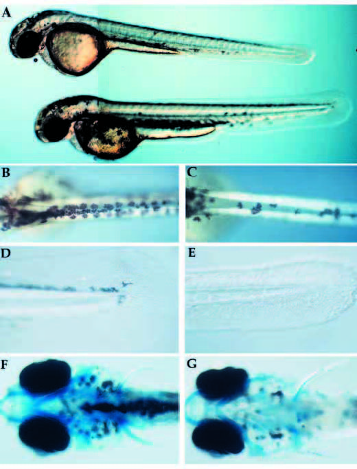
Fig. 3
- ID
- ZDB-IMAGE-140317-16
- Publication
- Artinger et al., 1999 - Zebrafish narrowminded suggests a genetic link between formation of neural crest and primary sensory neurons
- All Figures
- Figures for Artinger et al., 1999
Image
|
Figure Caption
Fig. 3 Visible phenotype of nrd and defects in formation of neural crest derivatives. (A) Whole live nrd (top) and wild-type sibling (bottom) at 48 hpf (lateral view). Wild-type (B,D,F) and nrd (C,E,G) siblings (10x). nrd mutant larvae have a smaller number of pigment cells (B,C: dorsal view), reduced fin mesenchyme (D,E: lateral view) and a normal pattern of cartilage (F,G: ventral view, visualized in whole mount by alcian blue stain. The slight reduction of density of the stain is not observed in serial sections).
Figure Data
Acknowledgments
This image is the copyrighted work of the attributed author or publisher, and
ZFIN has permission only to display this image to its users.
Additional permissions should be obtained from the applicable author or publisher of the image.
Full text @ Development