
Fig. 6
- ID
- ZDB-IMAGE-140314-1
- Publication
- Chandrasekhar et al., 1999 - The zebrafish detour gene is essential for cranial but not spinal motor neuron induction
- All Figures
- Figures for Chandrasekhar et al., 1999
|
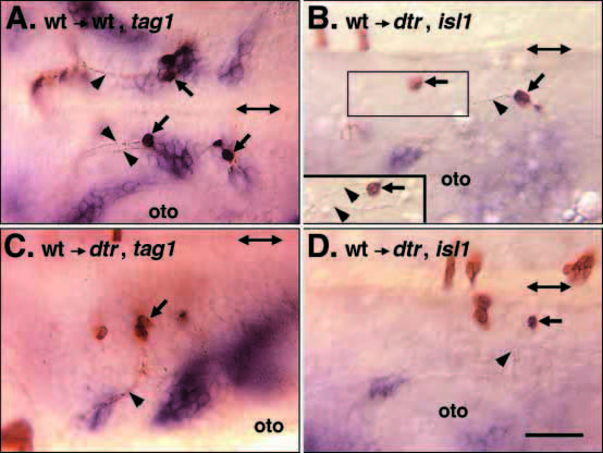
Fig. 6 detour functions cell autonomously in branchiomotor neuron induction. All panels show dorsal views of the hindbrain, with rostral to the left, in whole-mounted embryos processed for tag1 (A,C) or islet1 (B,D) in situ hybridization. Double arrows in each panel mark the midline. Donor wildtype cells and their axons are labeled brown. Embryos in B-D were identified as mutant host embryos because their hindbrains did not contain the characteristic clusters of branchiomotor neurons expressing tag1 (C) or islet1 (B,D). (A) When wild-type cells are transplanted into a wild-type host embryo, the donor cells within the nVII neuronal clusters (arrows) extend axons (arrowheads) anteriorly that exit the hindbrain in r4 into the hyoid arch, as described previously (Chandrasekhar et al., 1997). (B) In a dtr host embryo, donor wild-type cells (arrows) in r4 and r6 extend axons (arrowhead) anteriorly. The boxed area is depicted in a different focal plane (inset) to show that the axons (arrowheads) turn laterally exiting the hindbrain in r4 into the hyoid arch, in a manner characteristic of nVII axons. (C) In another dtr host embryo, donor wild-type cells (arrow) in r2 extend axons (arrowhead) laterally that exit the hindbrain in r2 into the mandibular arch, in a manner characteristic of nV axons. (D) In a third dtr host embryo, a donor wild-type cell (arrow) in r6 extends an axon (arrowhead) laterally that exits the hindbrain in r6, in a manner characteristic of nIX axons. oto, otocyst. Scale bar, 40 μm.