
|
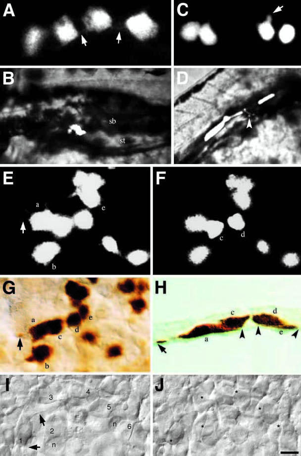
Fig. 5 Endodermal precursors occupy a stereotypic depth in the hypoblast. (A) Flattened cells, characteristic of the endodermal precursors in the late gastrula; note faint filopodia (arrows). At 100% epiboly, these cells were located directly adjacent to the yolk cell. (B) By 96 hours, these cells had differentiated into swimbladder tissue (sb) neighboring the stomach (st). (C) Rounded cells, characteristic of mesodermal precursors in the late gastrula; note lack of filopodia, however some cells show protrusive activity (arrow). At 90% epiboly, these cells were intermingled within a coherent layer of unlabeled cells of similar morphology one cell diameter up from the yolk cell. (D) By 30 hours, these cells had differentiated into paraxial muscle and 1 endothelial cell (arrow). (E-H) Five clones were examined at 90% epiboly and then immediately fixed, stained and sectioned. Illustrated is one example. The clone in the live embryo, deep (E) and superficial (F) planes of focus. Cells a, b and e are large and flattened endodermal precursors; one of several filopodia on cell a is indicated (arrow). Cells c and d are smaller and rounder mesodermal precursors, note cell c partially overlies cell a. (G) The same cohort of cells in the whole-mount preparation; note the filopodia on cell a (arrow). (H) Transverse section (10 mm) through cells a, c, d, and e. Both cell a and e are in the deepest hypoblast cell layer, beneath cells c and d. The extremities of cell a and e are indicated with arrowheads and an arrow, which further indicates the filopodia on cell a. (I,J) Unlabeled cells in the live embryo at 100% epiboly. The field of view is located 45° away from the dorsal midline near the level of the equator, because the embryo is spherical, cells at the bottom of the field are in a deeper plane of focus than cells at the top. (I) Deep plane of focus, six endodermal precursor cells (indicated with numerals) – each cell spaced roughly one cell diameter apart from one another – overlay the yolk cell whose nuclei (n) are also in focus. Filopodia (arrows) on cell 1 are indicated. (J) Superficial plane of focus, a layer of mesodermal precursor cells lie above the endodermal cells. Mesodermal precursor cells form a coherent layer – there are no spaces between cells. Asterisk denotes the positions of the underlying endoderm cells. Notice cells 1, 4 and 5 are partially visible. Scale bar: 10 μm (A,C, E-G, and I,J), 50 μm (B,D), 5 μm (H).