
Image
|
Figure Caption
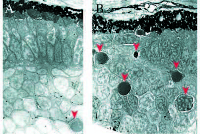
Fig. 8 Electron micrographs of frontal sections from retinae at 76 hpf. (A) Wild-type retina; retinal pigmented epithelium is to the top. Note the already formed PR cell layer with outer segments. (A) Single apoptotic cell can be seen (arrowhead). (B) nrf/nrf sibling, note the discontinuous PR layer and high numbers of apoptotic profiles (arrowheads).
Figure Data
Acknowledgments
This image is the copyrighted work of the attributed author or publisher, and
ZFIN has permission only to display this image to its users.
Additional permissions should be obtained from the applicable author or publisher of the image.
Full text @ Development