
Fig. 7
- ID
- ZDB-IMAGE-140307-40
- Antibodies
- Publication
- Haddon et al., 1998 - Delta-Notch signaling and the patterning of sensory cell differentiation in the zebrafish ear: evidence from the mind bomb mutant
- All Figures
- Figures for Haddon et al., 1998
|
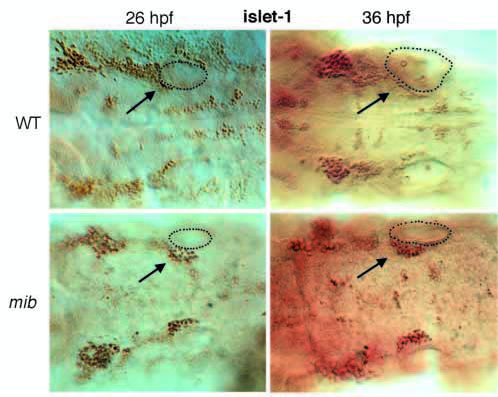
Fig. 7 Dorsal view of ears plus hindbrain at 26 and 36 hpf, stained with islet-1 antibody using HRP detection, showing the statoacoustic ganglion (arrows) containing many more neurons in mib than in wild type. The outline of the right ear is indicated by a dotted line. The statoacoustic ganglion at early stages forms a continuous mass with the anterior lateral line ganglion and the VIIth cranial nerve ganglion. As a rough guide, however, one can take the statoacoustic ganglion cells to be those that lie at and posterior to the anterior end of the otocyst; we followed this rule in making the counts described in the text.