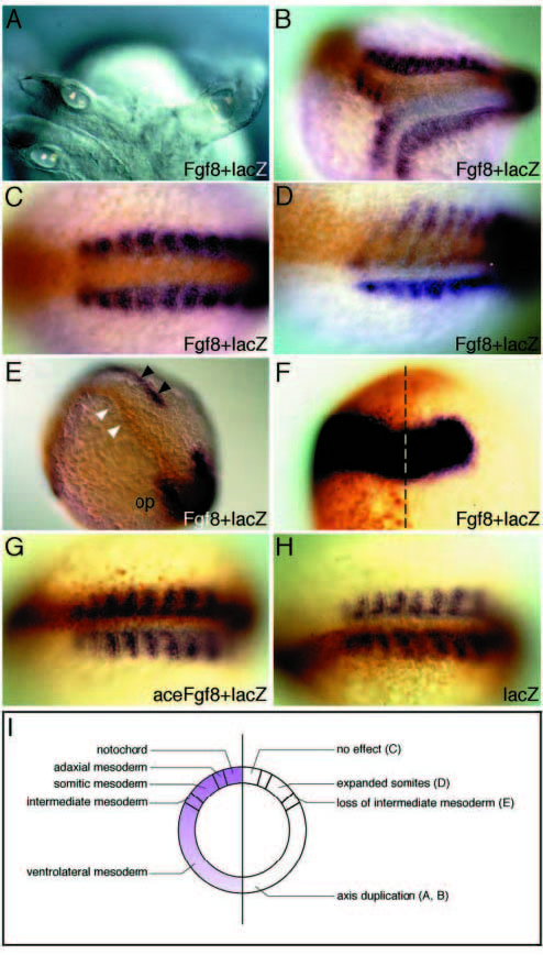
Fig. 6 Function of wild-type and mutant Fgf8 in dorsoventral patterning of the gastrula. Adaxial and somitic mesoderm is visualized with myoD (blue), or intermediate mesoderm and MHB with Pax2.1, and location of the lacZ co-injected cells with an antibody to β-gal (brown). RNA distribution is mostly restricted to one side, allowing comparison with the contralateral side as a control. (A-F) Misexpression of Fgf8 in wild-type embryos by RNA injection. (A,B) Live 28 h embryo with axis duplication (A) and stained for myoD (B). (C) Axial location of the injected RNA yields no obvious defect in mesoderm patterning. (D) Expansion of somitic mesoderm on the injected side of embryo. (E) Pax2.1-positive intermediate mesoderm (black arrowheads) is missing on the injected side (white arrowheads). (F) Fgf8 misexpression alters the d/v, but not the a/p extent of the Pax2.1 expression domain on the left, injected side (midline is given by a dashed line; op, otic placode). (G) No effect is seen after injection of the acerebellar version (lacking exon2) of Fgf8 RNA, showing that the acerebellar transcript is inactive in vivo. (H) lacZ control injections had no effect. (I) Summary of the effects observed after injection of Fgf8. Left: schematic fate map of a gastrula, and the Fgf8 gradient in the germ ring. Right: consequence of misexpressing Fgf8 in the respective area. All embryos shown are at early to midsomitogenesis stages; BD, G, H show myoD in situ stainings of injected embryos, E and F show Pax2.1 in situ stainings; β-gal was detected by antibody staining.
Image
Figure Caption
Acknowledgments
This image is the copyrighted work of the attributed author or publisher, and
ZFIN has permission only to display this image to its users.
Additional permissions should be obtained from the applicable author or publisher of the image.
Full text @ Development

