
Fig. 1
- ID
- ZDB-IMAGE-140304-67
- Antibodies
- Publication
- Chen et al., 1996 - Mutations affecting the cardiovascular system and other internal organs in zebrafish
- All Figures
- Figures for Chen et al., 1996
|
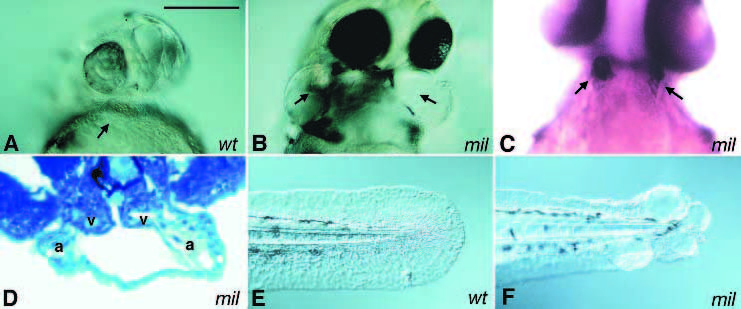
Fig. 1 Cardia bifida mutants. Two hearts are located at either side of the embryo. A ventral view of 1-day old embryos shows two swollen pericardial sac in milte273 (B) but not in the wild-type sibling (A). Two hearts can be clearly visualized either by immunostaining or in histological sections. (C) A 2-day old milte273 embryo stained with the antibody S46, which labels the atria. (D) A transverse section of a 2-day old milte273 embryo. The hearts are indicated by arrows; a, atrium; v, ventricle. Images of tails of live 1-day old wild-type and milte273 embryos are shown in E and F, respectively. Scale bar, 100 μm.