
IMAGE
Fig. S1
- ID
- ZDB-IMAGE-140304-39
- Publication
- He et al., 2014 - Maternal control of axial-paraxial mesoderm patterning via direct transcriptional repression in zebrafish
- All Figures
- Figures for He et al., 2014
Image
|
Figure Caption
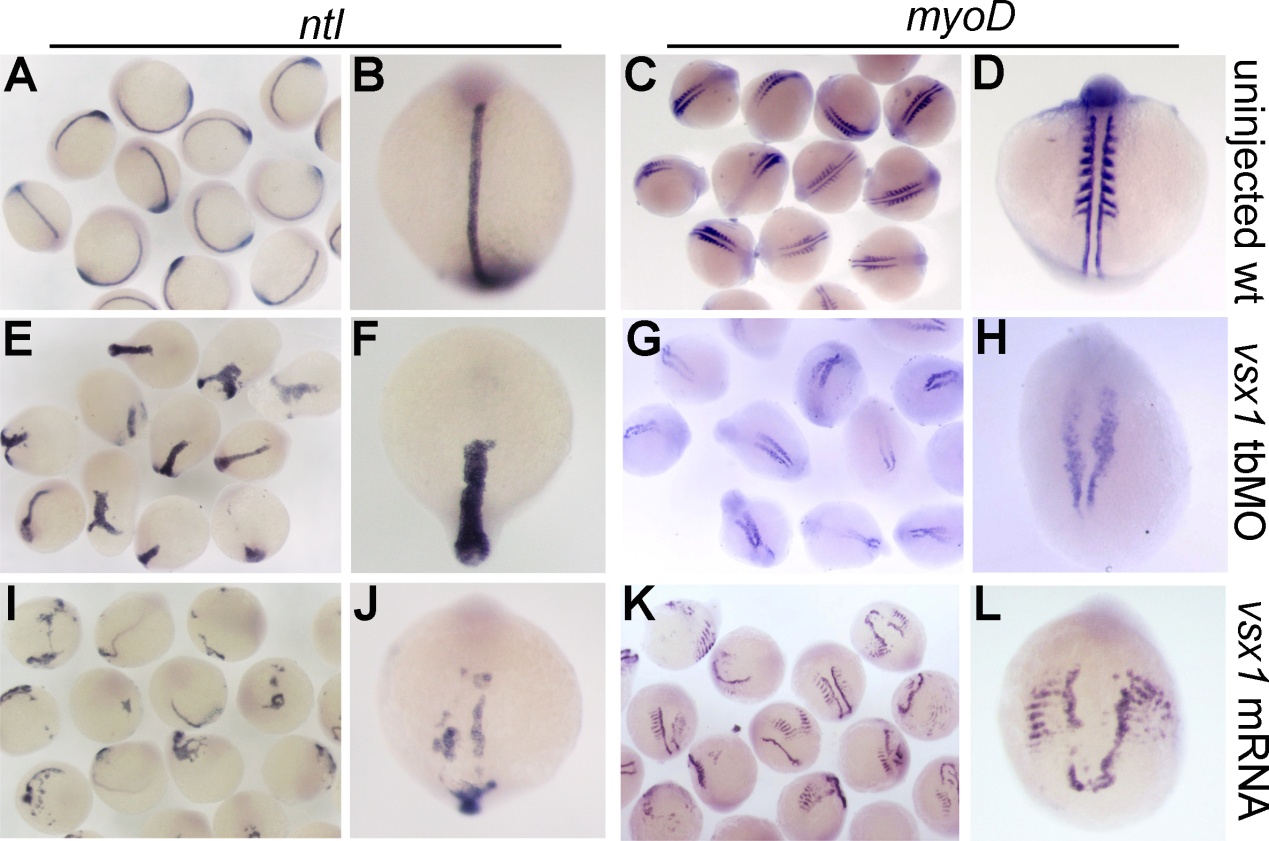
Fig. S1
Comparison of axial and paraxial mesoderm formation in uninjected wildtype control, vsx1 knockdown and overexpression embryos at 8-10 somite stage. (A-B, E-F, I-J) Axial mesoderm domain is visualized by whole mount in situ hybridization with marker gene ntl. (C-D, G-H, K-L) Paraxial mesoderm domain is visualized by whole mount in situ hybridization with marker gene myoD.
Acknowledgments
This image is the copyrighted work of the attributed author or publisher, and
ZFIN has permission only to display this image to its users.
Additional permissions should be obtained from the applicable author or publisher of the image.
Reprinted from Developmental Biology, 386(1), He, Y., Xu, X., Zhao, S., Ma, S., Sun, L., Liu, Z., and Luo, C., Maternal control of axial-paraxial mesoderm patterning via direct transcriptional repression in zebrafish, 96-110, Copyright (2014) with permission from Elsevier. Full text @ Dev. Biol.