
|
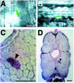
Fig. 9 Lateral presomitic cells become fast muscle fibers. (A) Dorsal view of the segmental plate of an approximately 15.5 h embryo (13 somites), after injection of lateral presomitic cells with lysinated fluorescein dextran. (B) Side view of the same embryo at about 38 h of development. One of the lateral presomitic cells developed into a ventral muscle fiber located in somite 15. (C) Transverse section of the same embryo at 15 d. The injected lateral presomitic cell developed into a large diameter muscle fiber (arrowhead) located deep in the muscle. (D) Lower magnification view of the injected cell (arrowhead). The muscle fiber is within the fast muscle region. In a series of similar experiments, 3 out of 3 injected lateral presomitic cells became fast muscle fibers. In dorsal views, anterior is up; in side views, anterior is to the left; in transverse sections, dorsal is up. Scale bars, 50 μm.