
IMAGE
Fig. 7
- ID
- ZDB-IMAGE-140228-29
- Publication
- Schilling et al., 1996 - The chinless mutation and neural crest cell interactions in zebrafish jaw development
- All Figures
- Figures for Schilling et al., 1996
Image
|
Figure Caption
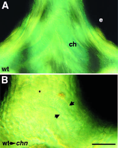
Fig. 7 Rescue of cartilage patterning in chn by transplanted wildtype neural crest cells. Nomarski images of cartilage. 72 hour, ventral view, anterior to the top. (A) Alcian-blue-stained cartilage in the bilateral ceratohyals of a wild-type embryo. (B) A mosaic embryo showing a rod of cells with the stacked appearance of chondrocytes (between arrowheads), extends to the ventral midline in the position of the ceratohyal (asterisk). Abbreviations: ch, ceratohyal; e, eye. Scale bars, 100 μm.
Acknowledgments
This image is the copyrighted work of the attributed author or publisher, and
ZFIN has permission only to display this image to its users.
Additional permissions should be obtained from the applicable author or publisher of the image.
Full text @ Development