
Image
|
Figure Caption
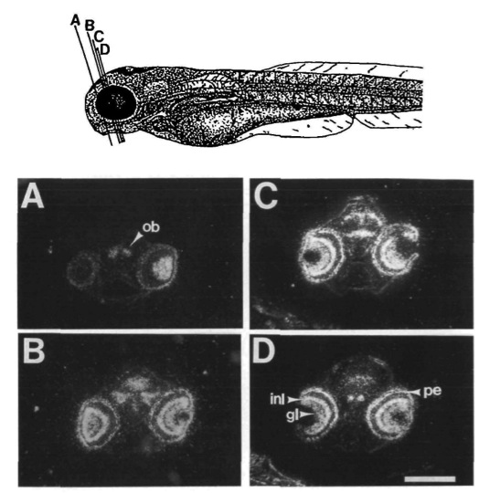
Fig. 8 pax-6 is still expressed in the head at hatching (72 h). Cross-sections of 3-day fish were hybridized with pax-6 specific probes. Dorsal is up. The sketch in the upper part indicates approximate levels of the sections. Expression is apparent in the olfactory bulb (A, ob), the ganglion cell layer (gl) and the inner nuclear layer (inl) of the eye (A - D). Specific parts of the diencephalon express pax-6 (C - D). The signal in the pigment epithelium (pe) of the eye is due to pigment and does not represent a hybridization signal. Scale bar is 200 μm in A - D and 290 μm in drawing.
Figure Data
Acknowledgments
This image is the copyrighted work of the attributed author or publisher, and
ZFIN has permission only to display this image to its users.
Additional permissions should be obtained from the applicable author or publisher of the image.
Full text @ Development