
Fig. 6
- ID
- ZDB-IMAGE-140228-12
- Genes
- Publication
- Püschel et al., 1992 - Sequence and expression pattern of pax-6 are highly conserved between zebrafish and mice
- All Figures
- Figures for Püschel et al., 1992
|
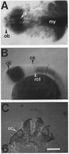
Fig. 6 pax-6 is expressed in the brain and spinal cord. (A) 24 h embryo. Expression is seen in the olfactory bulb (ob), the diencephalon (di), the eye and the myelencephalon (my). (B) Expression in the hindbrain starts at the level of the first rhomobomere (rol). The line indicates the level of the cross-section in C. (C) A double-exposure of the bright-field (blue) and dark-field (red) images of the same cross-section of the hindbrain at the level of the otocyst (oc). The domain of pax-6 expression is seen as a red signal. Expression in the hindbrain is restricted to the ventral half excluding the floorplate. Anterior is to the left in A and B and dorsal is up. Scale bar is 100 μm in A; 200 μm in B; and 50 μm in C.