
Fig. 4
- ID
- ZDB-IMAGE-140221-6
- Publication
- Drummond et al., 1998 - Early development of the zebrafish pronephros and analysis of mutations affecting pronephric function
- All Figures
- Figures for Drummond et al., 1998
|
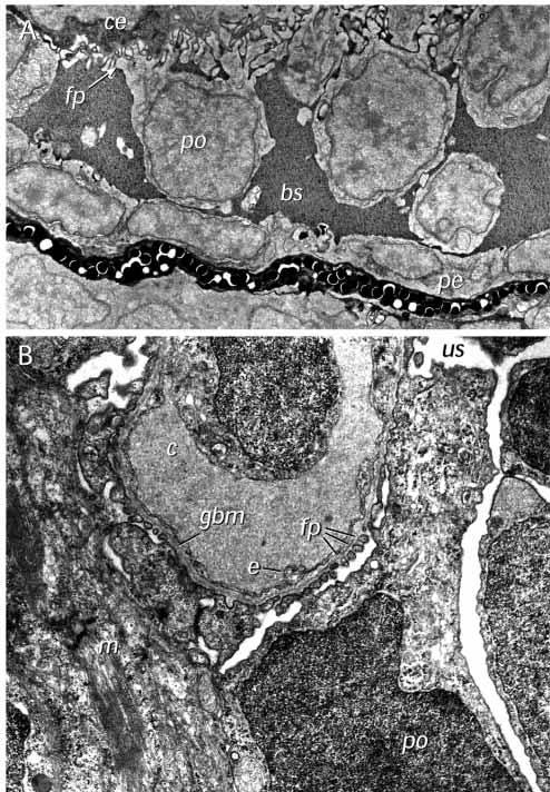
Fig. 4 Electron microscopy of the wild-type glomerulus. (A) Ultrastructure of the forming glomerulus at 40 hpf. A longitudinal section (similar to that presented in Fig. 2D) shows podocytes (po) extending foot processes (fp) in a dorsal direction and in close contact with capillary endothelial cells (ce). Bowman’s space (bs) appears filled with an electron-dense precipitate and the ventralmost cells appear to be taking on the flattened appearance of the parietal epithelium (pe) that will line Bowman’s capsule. (B) Glomerulus and filtration barrier of the wild-type pronephros at 3.5 days postfertilization. The filtration barrier is well developed displaying an endothelium with open pores (without diaphragm), a thin trilaminar glomerular basement membrane (gbm) and podocytes with primary processes and numerous interdigitating foot processes (fp). The filtration slits between the foot processes are bridged by one or sometimes two slit membranes. Mesangial cells (m) are also evident. us, urinary space; c, capillary space; e, endothelial cell. ×36,500.