
Fig. 4
- ID
- ZDB-IMAGE-140117-28
- Publication
- Ryan et al., 2013 - Rapid identification of kidney cyst mutations by whole exome sequencing in zebrafish
- All Figures
- Figures for Ryan et al., 2013
|
Fig. 4
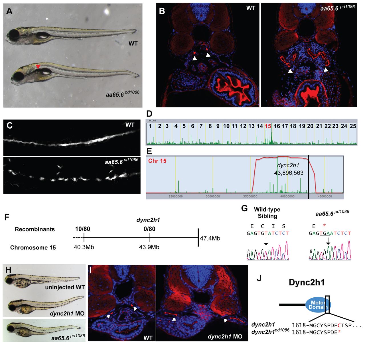
A truncation at the end of the motor domain of Dync2h1 leads to a cystic kidney phenotype. (A) Brightfield image of 5-dpf WT sibling and aa65.6pd1086 mutant larvae. Red arrowhead indicates a kidney cyst. (B) Confocal images of transverse sections of 5-dpf WT sibling and aa65.6pd1086 mutant larvae stained with phalloidin (red) and DAPI (blue). The arrowheads point to pronephric ducts and asterisks mark kidney cysts. (C) Confocal image of 3-dpf WT and aa65.6pd1086 mutant embryos in whole-mount, stained for acetylated tubulin to mark cilia. (D) Linkage analysis for aa65.6pd1086 using SNPtrack. (E) The homozygosity interval for aa65.6pd1086. (F) Linkage analysis using single embryos. (G) Sequencing of genomic DNA of WT and aa65.6pd1086 mutant larvae. (H) Injection of a morpholino directed against a splice site within the motor domain of dync2h1 phenocopies the aa65.6pd1086 mutation. (I) Confocal images of transverse sections 3-dpf WT and dync2h1 morphants stained with phalloidin and DAPI, showing distended pronephric ducts (arrowheads). (J) Schematic of the Dync2h1 protein illustrating that the mutation removes a portion of the motor domain.