
Fig. S4
- ID
- ZDB-IMAGE-140117-12
- Publication
- Nagashima et al., 2013 - A self-renewing division of zebrafish Muller glial cells generates neuronal progenitors that require N-cadherin to regenerate retinal neurons
- All Figures
- Figures for Nagashima et al., 2013
|
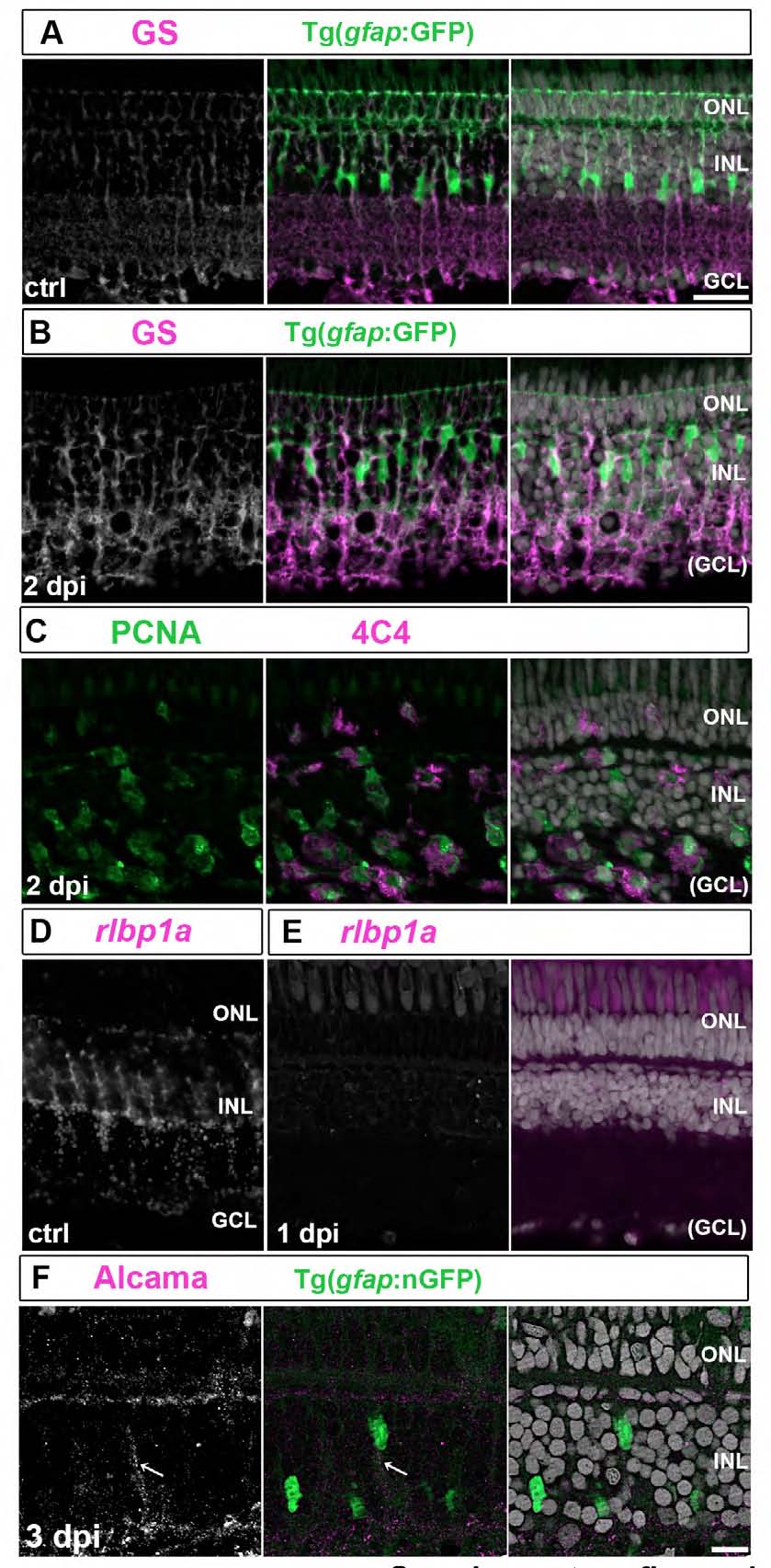
Fig. S4
Basal processes of Muller glia collapse and markers of differentiation are downregulated after intraocular ouabain injection.
(A, B) Immunocytochemistry for the Müller glial marker glutamine synthetase (GS) (white/magenta) in unlesioned (A) and 2 dpi (B) gfap:GFP retinas with Müller glial reporter (green). GS+/GFP+ Müller glial processes in the inner retina are disrupted, whereas the organization of the outer retina, including the adherens junctions at the outer limiting membrane, remain intact at 2 dpi. (C) PCNA+ (green) activated microglia (4C4+, magenta) are abundant in the damaged inner retina at 2 dpi. (D, E) In situ hybridization for another Müller glia marker, rlbp1a (white/magenta, D), shows expression is gone at 1 dpi (E). (F) Weak Alcama immunoreactivity (magenta/ white) appears in the basal process (arrow) of a Müller glia with the inducible nGFP reporter (green) at 3 dpi. Scales: 20 μm, A-E; 10 μm, F.