
|
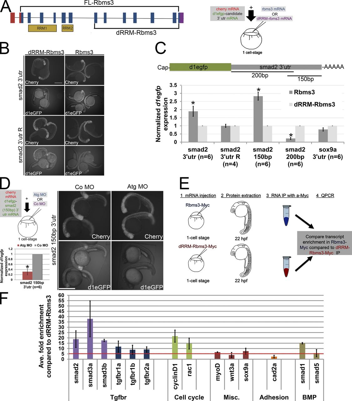
Fig. 8 Smad2 transcripts are a target of Rbms3. (A) dRRM-Rbms3 lacks the putative RNA-binding domains (RRM; lower bracket, left). A schematic showing an in vivo stability assay is shown on the right. (B and C) Fluorescent images (B) and QPCR (C; d1egfp normalized to cherry) showing that full-length Rbms3 treatment stabilizes d1egfp-smad2 32 utr and d1egfp-smad2 (150bp) 32 utr reporters. (D) Schematic shows the experiment (top left), QPCR (d1egfp normalized to cherry; bottom left), and fluorescent images (right) showing that Atg MO treatment destabilizes d1egfp-smad2 (150bp) 32 utr. (B and D) Images were taken under same brightness/contrast conditions. (E) Schematic of RIP experiment. (F) QPCR showing transcripts enriched (more than fivefold, red dotted line) for full-length Rbms3 (n = 2–3 experiments). Error bars indicate SEM (C) and SD (D and F). n = RNA from individual embryos (C and D). *, P < 0.05 (Student’s t test). Bars, 500 µM.