
|
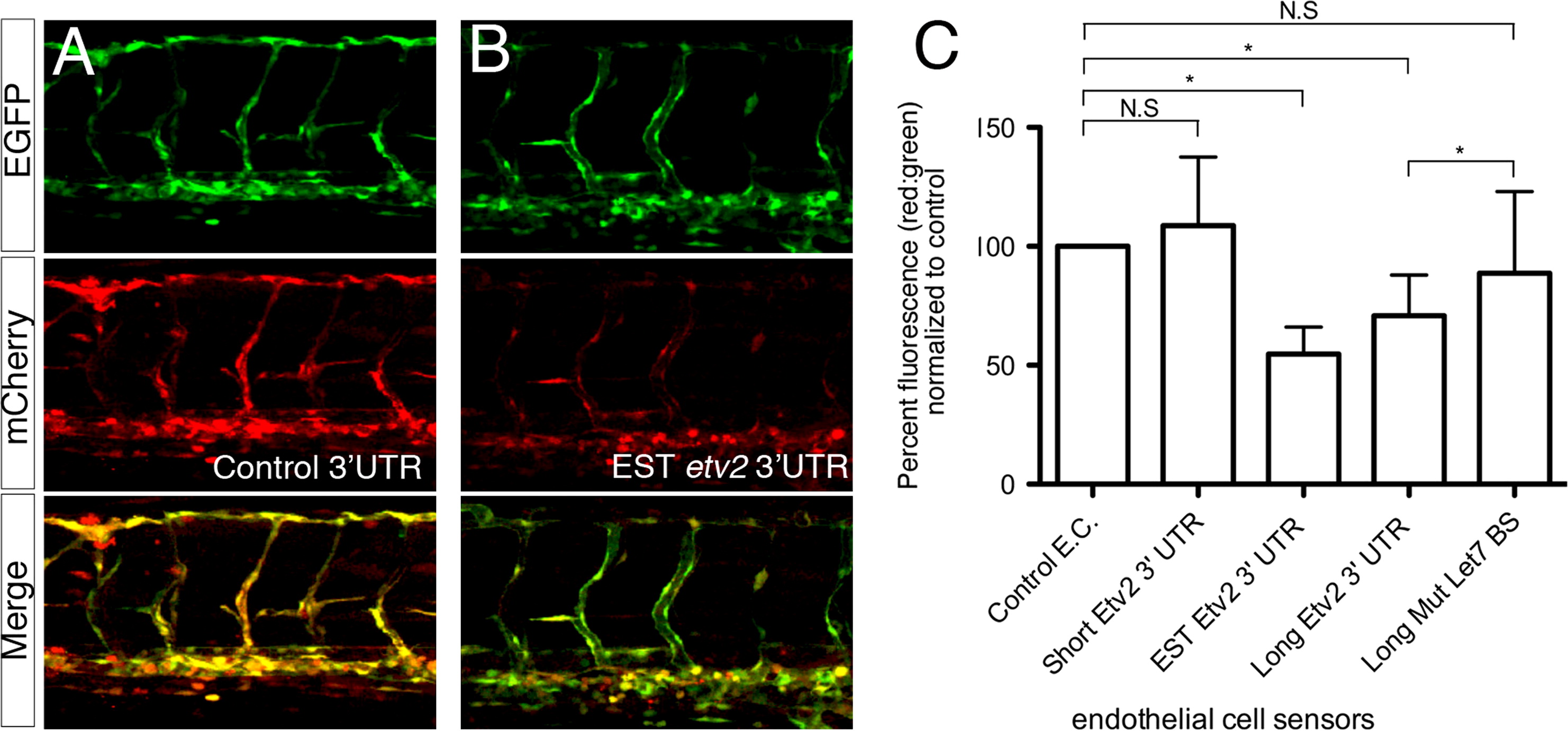
Fig. 3 The etv2 3′UTR represses a heterologous reporter in endothelial cells. (A, B) Representative confocal micrographs of 48 hpf wild type embryos co-injected with 25 pg of transposase mRNA and 25 pg of a Tol2 bis-cistronic endothelial cell autonomous sensor construct encoding mCherry fused to a (A) control 3′UTR or the (B) EST etv2 3′UTR sensor. (Top) Endothelial expression of the control EGFP transgene. (Middle) Endothelial expression of the mCherry sensor transgene. (Bottom) Merge of green and red channels. Lateral views, dorsal is up, anterior to the left. (C) Quantification of relative mCherry fluorescence levels compared to EGFP following 3′ UTR sensor injection. *p<0.05, N. S.=Not significant.
Reprinted from Developmental Biology, 384(1), Moore, J.C., Sheppard, S., Shestopalov, I.A., Chen, J.K., and Lawson, N., Post-transcriptional mechanisms contribute to Etv2 repression during vascular development, 128-40, Copyright (2013) with permission from Elsevier. Full text @ Dev. Biol.