
Image
|
Figure Caption
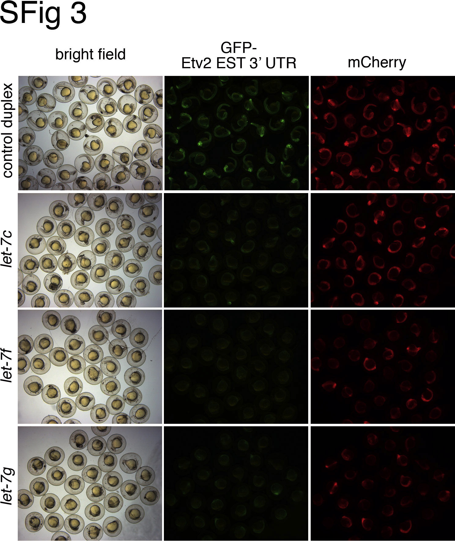
Fig. S3 Multiple let-7 family members can repress the etv2 3′ UTR. Embryos were co-injected with gfp-est-etv2 3′ UTR sensor (25 pg) and mcherry mRNAs (25 pg), along with indicated RNA duplexes. Bright field (left column), green fluorescent (middle column) and red fluorescent (right column) images of injected embryos were captured at 24 hpf.
Acknowledgments
This image is the copyrighted work of the attributed author or publisher, and
ZFIN has permission only to display this image to its users.
Additional permissions should be obtained from the applicable author or publisher of the image.
Reprinted from Developmental Biology, 384(1), Moore, J.C., Sheppard, S., Shestopalov, I.A., Chen, J.K., and Lawson, N., Post-transcriptional mechanisms contribute to Etv2 repression during vascular development, 128-40, Copyright (2013) with permission from Elsevier. Full text @ Dev. Biol.