
IMAGE
Fig. S2
- ID
- ZDB-IMAGE-131231-39
- Publication
- van der Velden et al., 2013 - The polycomb group protein ring1b/rnf2 is specifically required for craniofacial development
- All Figures
- Figures for van der Velden et al., 2013
Image
|
Figure Caption
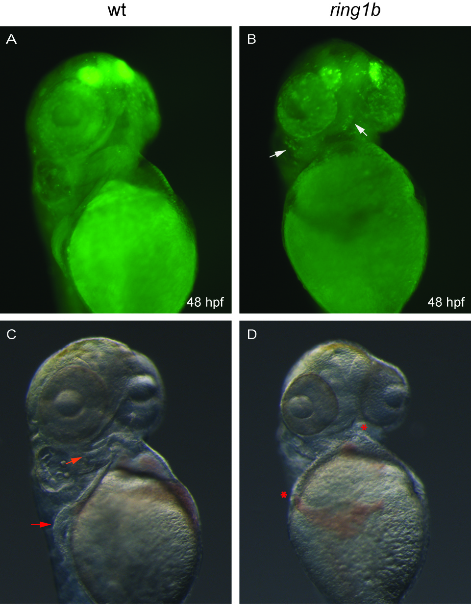
Fig. S2 Persistence of apoptotic cells in ring1b mutants. Ventral views of WT and ring1b embryos at 48 hpf stained with Acridine Orange. No apoptotic cells are detected in the pharyngeal arch region of WT embryos (A) whereas few AO-positive apoptotic cells have persisted in the ring1b mutants (B) Arrows in (B) indicate apoptotic clusters in the prospective jaw region and anteriorly to the otic vesicle. Figure also shows images of live WT and ring1b embryos depicting the fin and pharyngeal cartilages in WT (C, arrows) and their absence in the ring1b mutants (D, *).
Acknowledgments
This image is the copyrighted work of the attributed author or publisher, and
ZFIN has permission only to display this image to its users.
Additional permissions should be obtained from the applicable author or publisher of the image.
Full text @ PLoS One