Image
Figure Caption
Fig. 4
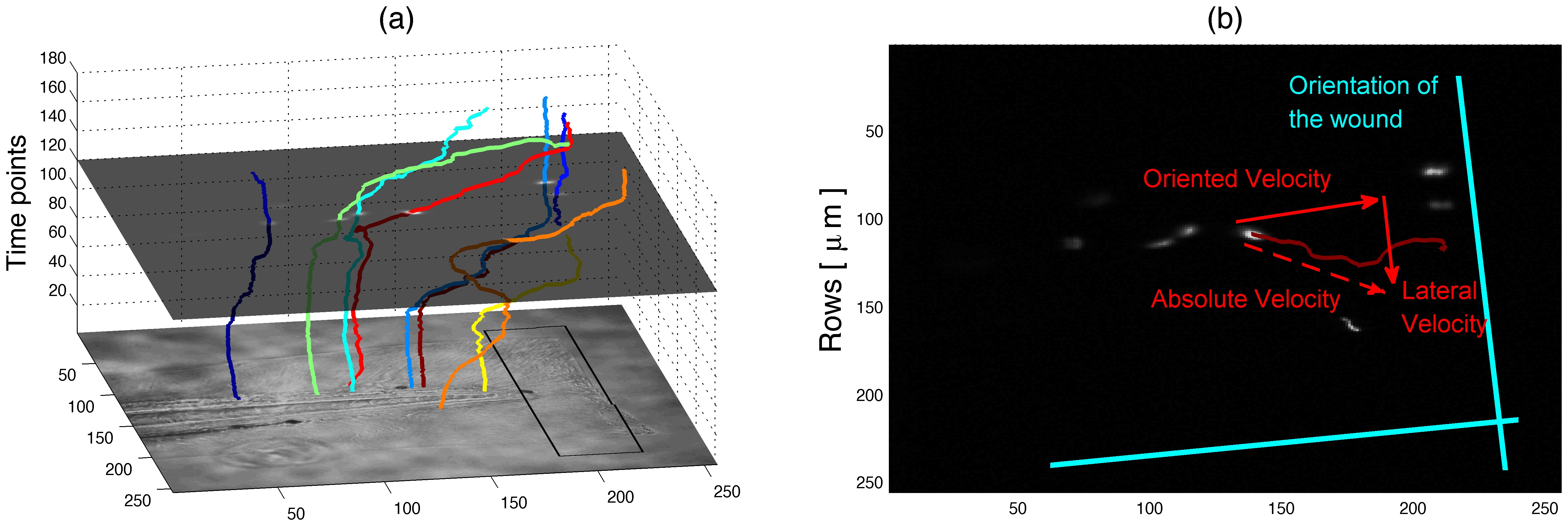
Orientation of the movement based on the artificial wound region.
(a) Three-dimensional plot of the tracks with time as the vertical axis. The DIC image of the fish is presented as a horizontal plane at time 0 and one fluorescent slice is shown at time 120. The black square over the DIC corresponds to the artificial wound region. (b) Description of the absolute, oriented and lateral neutrophil velocities with respect to the axis defined according to a manual delineation of the wound region.
Acknowledgments
This image is the copyrighted work of the attributed author or publisher, and
ZFIN has permission only to display this image to its users.
Additional permissions should be obtained from the applicable author or publisher of the image.
Full text @ PLoS One

