
IMAGE
Fig. S3
- ID
- ZDB-IMAGE-131213-36
- Publication
- Lam et al., 2013 - odd-skipped related 2 is required for fin chondrogenesis in zebrafish
- All Figures
- Figures for Lam et al., 2013
Image
|
Figure Caption
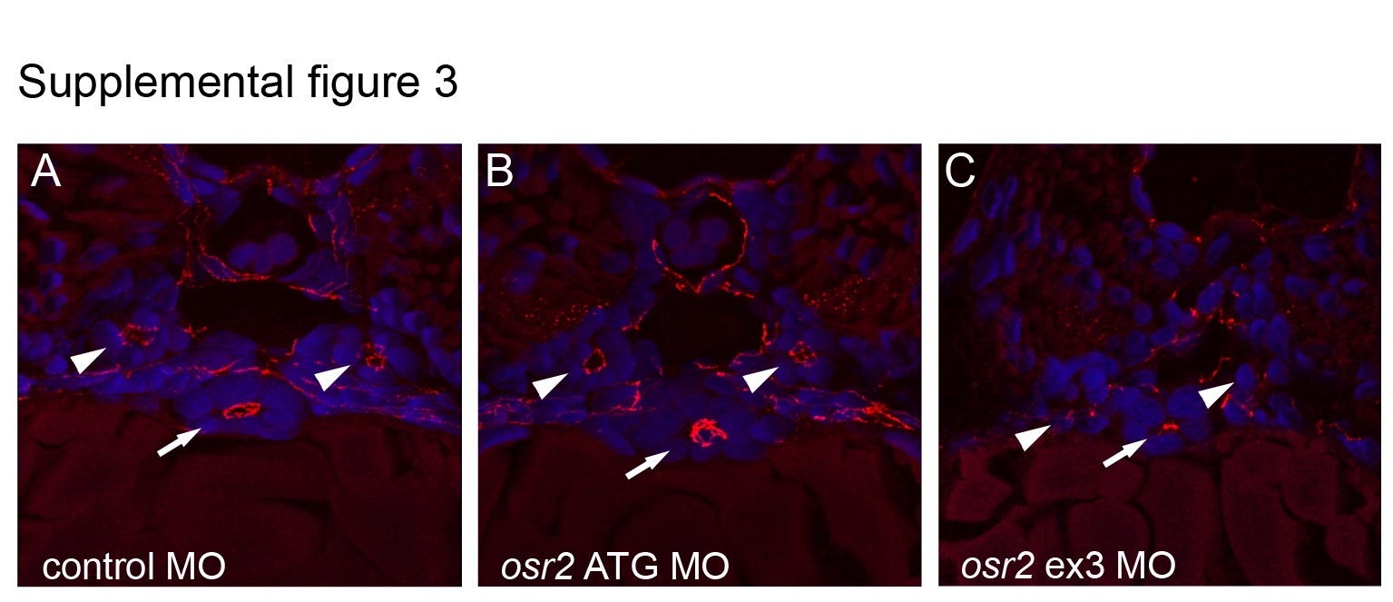
Fig. S3 Epithelial development and polarity is not affected in osr2 morphants. A: Histological section of a control embryo at the level of the mid-trunk shows cross-sections of the kidney (arrowheads) and gut (arrow) stained with anti-ZO-1, highlighting polarized apical cell–cell junctions. Both osr2 ATG morphants (B) and osr2 exon3 splice morphants (C) also show normal epithelial polarized junctions in the kidney and gut. Blue, DAPI stained nuclei. Representative sections from n=3 for each morpholino.
Acknowledgments
This image is the copyrighted work of the attributed author or publisher, and
ZFIN has permission only to display this image to its users.
Additional permissions should be obtained from the applicable author or publisher of the image.
Full text @ Dev. Dyn.