
IMAGE
Fig. 1
- ID
- ZDB-IMAGE-131211-42
- Genes
- Publication
- Kwon et al., 2013 - The parallel growth of motoneuron axons with the dorsal aorta depends on Vegfc/Vegfr3 signaling in zebrafish
- All Figures
- Figures for Kwon et al., 2013
Image
|
Figure Caption
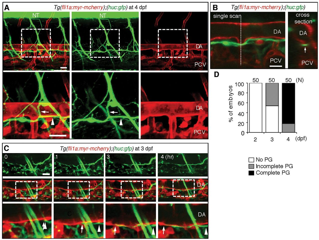
Fig. 1 Filopodia facilitate anastomosis of DLAV and the vein plexus. (A,B) Endothelial tip cells of untreated (A) and 0.08 μg/ml Lat. B-treated (B) 31 hpf Tg(Fli1ep:Lifeact-EGFP); Tg(Kdr-l:ras-Cherry)s916 zebrafish embryos at the dorsal roof of the neural tube. Arrows indicate polymerisation of F-actin at new cell contacts. Time is in minutes. (C-D′) Vein plexi of Tg(Fli1ep:Lifeact-EGFP); Tg(Kdr-l:ras-Cherry)s916 embryos treated with DMSO or 0.1 μg/ml Lat. B from 31 to 46 hpf. Arrowheads indicate junctional F-actin. DA, dorsal aorta; DV, dorsal vein; VV, ventral vein. Scale bars: 10 μm.
Figure Data
Acknowledgments
This image is the copyrighted work of the attributed author or publisher, and
ZFIN has permission only to display this image to its users.
Additional permissions should be obtained from the applicable author or publisher of the image.
Full text @ Development