
Fig. 1
- ID
- ZDB-IMAGE-131210-21
- Genes
- Publication
- Zhen et al., 2013 - Hemogenic endothelium specification and hematopoietic stem cell maintenance employ distinct Scl isoforms
- All Figures
- Figures for Zhen et al., 2013
|
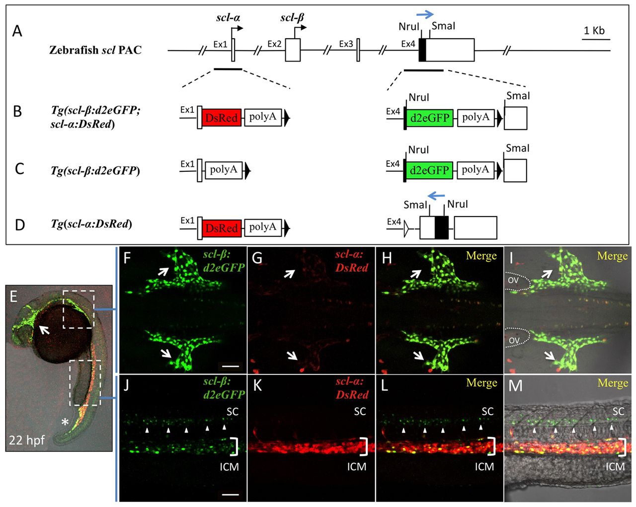
Fig. 1 Generation of scl transgenic lines and expression of d2eGFP and DsRed coincides with that of the endogenous scl-β and scl-α isoforms. (A) The structure of the zebrafish scl locus in the PAC clone BUSM-129I22. The transcription start sites of scl-α and scl-β are indicated (black arrows). The black box in exon 4 represents sequences encoding the basic helix-loop-helix (bHLH) domain. (B-D) The modified scl PAC used to generate Tg(scl-β:d2eGFP; scl-α:DsRed), Tg(scl-β:d2eGFP) and Tg(scl-α:DsRed). DsRed, an SV40 polyadenylation signal and a remaining FRT site were inserted behind exon 1 (B,D). d2eGFP, an SV40 polyadenylation signal and a remaining FRT site were inserted in exon 4 (B,C). The transcription activity of scl-α was interrupted by an SV40 polyadenylation signal immediately after exon 1 (C), whereas Scl-β expression was disrupted by the reversal of the protein coding sequences in exon 4 (D, blue arrow). (E-M) Expression of fluorescent reporter proteins in a 22 hpf Tg(scl-β:d2eGFP; scl-α:DsRed) embryo. Scl-β:d2eGFP is largely expressed in the head vasculature (E, arrow), the developing common cardinal vein (CCV) (F-I, arrows) and posterior part of the posterior blood island (PBI) (E, asterisk). By contrast, scl-α:DsRed is restricted in the intermediate cell mass (ICM) (J-M, bracket). (F-I) Higher magnification of d2eGFP and DsRed expression in the anterior lateral plate mesoderm region. D2eGFP is highly expressed in endothelial cells of the CCV across the yolk sac (arrows), whereas DsRed is almost undetected. (J-M) Higher magnification of d2eGFP and DsRed expression in the ICM region. DsRed is largely expressed in erythrocytes of the ICM, whereas d2eGFP is less expressed in ICM but dominant in neurons of the whole trunk segment of the spinal cord (arrowheads). The embryos are shown in lateral (E,J-M) or dorsal (F-I) views, with anterior to the left. ov, otic vesicle; sc, spinal cord. Scale bars: 50 μm.