
Fig. 2
- ID
- ZDB-IMAGE-131210-20
- Publication
- Hao et al., 2013 - Selective Small Molecule Targeting β-Catenin Function Discovered by In Vivo Chemical Genetic Screen
- All Figures
- Figures for Hao et al., 2013
|
Fig. 2
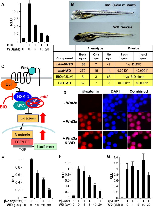
WD Blocks Wnt Signaling Downstream of β-Catenin Destruction Complex
(A) WD inhibited Wnt signaling induced by BIO in TOPFLASH-luciferase assays in STF293 cells (n = 4; results are represented as mean RLU ± SE).
(B) WD (20 μM) rescued the loss of telencephalon and eyes in mbl mutant zebrafish with defective Axin1 gene. Top view shows untreated Axin1/mbl mutant zebrafish embryo (3 days old). Bottom view presents WD-treated embryo with normal/restored eyes and telencephalon. Quantification of telencephalon/eye loss phenotype in mbl mutants and BIO-treated embryos, following treatment with WD or DMSO control, is shown. p value was determined using Student’s two-tailed t test.
(C) Model illustrates TOPFLASH-luciferase assay, with components of the Wnt/β-catenin signaling and various means to perturb them. Briefly, disruption of the β-catenin degradation complex, either through pharmacological inhibition of GSK3β using the small molecule BIO or a genetic mutation in Axin (mlb), results in nuclear β-catenin accumulation and subsequent Wnt reporter activation.
(D) WD treatment does not block Wnt3a-induced β-catenin nuclear translocation in RKO cells. RKO cells were immunostained for β-catenin (red) and counterstained with DAPI (blue) following 24 hr incubation without Wnt3a, with Wnt3a, and with Wnt3a plus WD (20 μM).
(E) WD inhibited Wnt signaling induced by overexpression of the constitutively active human β-catenin (cat) mutant (S33Y) in TOPFLASH-luciferase assay (n = 4; results are represented as mean RLU ± SE).
(F) WD blocked Wnt signaling induced by overexpression of the zebrafish β-catenin-1 in TOPFLASH-luciferase assay in STF293 cells (n = 3).
(G) WD did not block Wnt signaling induced by overexpression of the zebrafish β-catenin-2 (n = 3) in STF293 cells.
See also Figure S4.