
Fig. 2
- ID
- ZDB-IMAGE-131210-2
- Publication
- Sheehan-Rooney et al., 2013 - Ahsa1 and Hsp90 activity confers more severe craniofacial phenotypes in a zebrafish model of hypoparathyroidism, sensorineural deafness and renal dysplasia (HDR)
- All Figures
- Figures for Sheehan-Rooney et al., 2013
|
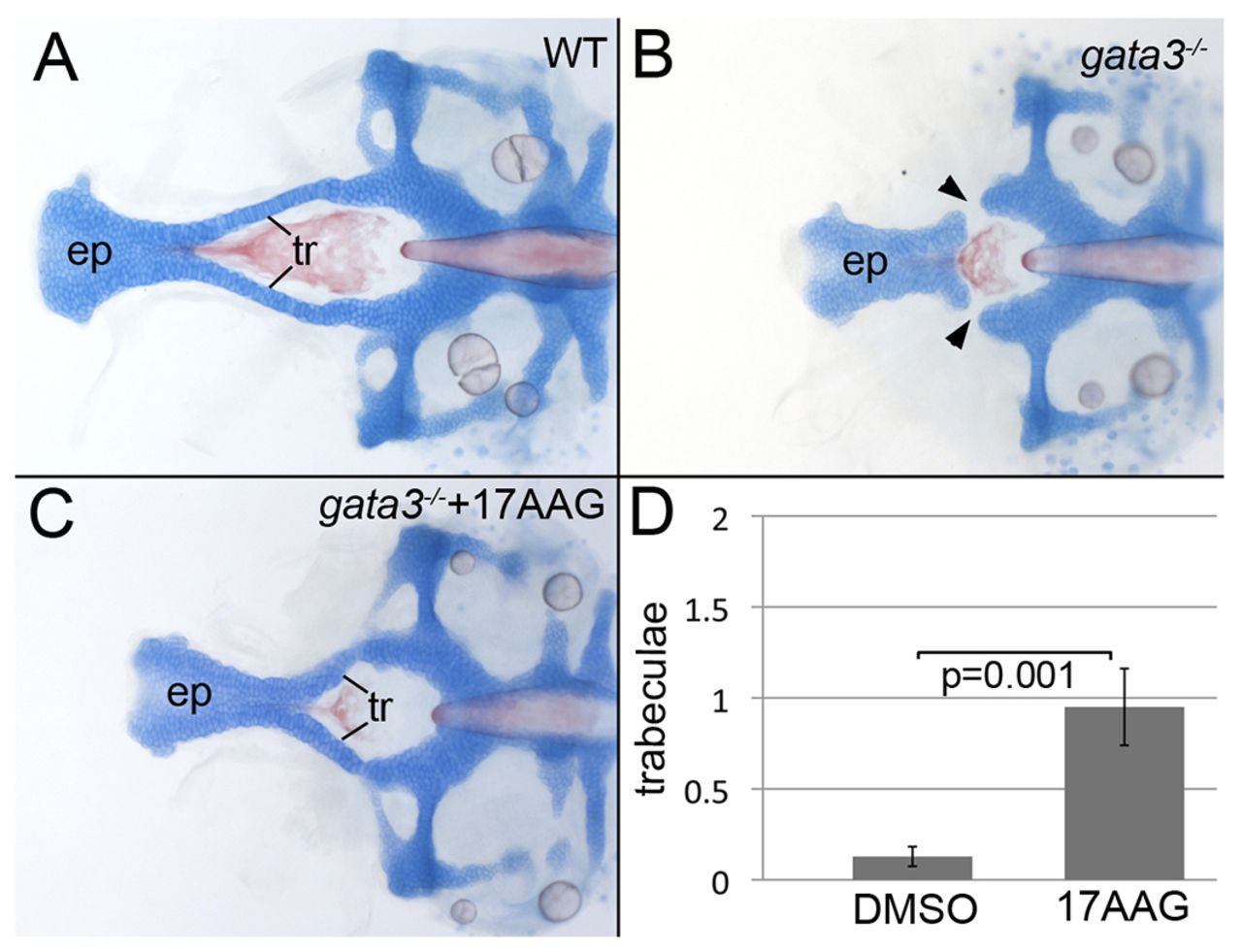
Fig. 2
Inhibition of Hsp90 partially restores trabeculae in severe mutants. (A) Zebrafish embryos were treated with levels of 17AAG that did not disrupt the craniofacial skeleton in wild type. (B) Untreated gata3 mutants in the severe background typically lack trabeculae (arrowheads). (C) Hsp90 inhibition partially restored the trabeculae, resulting in the fusion of the palate and posterior neurocranium. (D) 17AAG treatment of severe gata3 mutants significantly increases the number of trabeculae in treated embryos (average=0.95, s.e.m.=0.211, s.d.=0.94, n=20) compared with control, DMSO-treated, embryos (average=0.13, s.e.m.=0.05, s.d.=0.34, n=39). (A–C) Dorsal views of flat-mounted neurocrania, anterior to the left; ep, ethmoid plate; tr, trabeculae.