
IMAGE
Fig. S2
- ID
- ZDB-IMAGE-131108-19
- Genes
- Publication
- Choorapoikayil et al., 2013 - Loss of Pten promotes angiogenesis and enhanced vegfaa expression in zebrafish
- All Figures
- Figures for Choorapoikayil et al., 2013
Image
|
Figure Caption
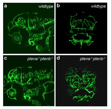
Fig. S2
Enhanced angiogenesis in head vasculature in ptena-/-ptenb-/- mutant embryos Endothelial cells in wildtype (a, b) and ptena-/-ptenb-/- mutants (c, d) were visualized using Tg(kdrl:eGFP) by confocal imaging at 4 dpf. Anterior to the left, 20x, 2 μm step size, lateral (a, c) and sagittal (b, d) view.
Figure Data
Acknowledgments
This image is the copyrighted work of the attributed author or publisher, and
ZFIN has permission only to display this image to its users.
Additional permissions should be obtained from the applicable author or publisher of the image.
Full text @ Dis. Model. Mech.