
IMAGE
Fig. 1
- ID
- ZDB-IMAGE-131108-13
- Genes
- Publication
- Choorapoikayil et al., 2013 - Loss of Pten promotes angiogenesis and enhanced vegfaa expression in zebrafish
- All Figures
- Figures for Choorapoikayil et al., 2013
Image
|
Figure Caption
Fig. 1
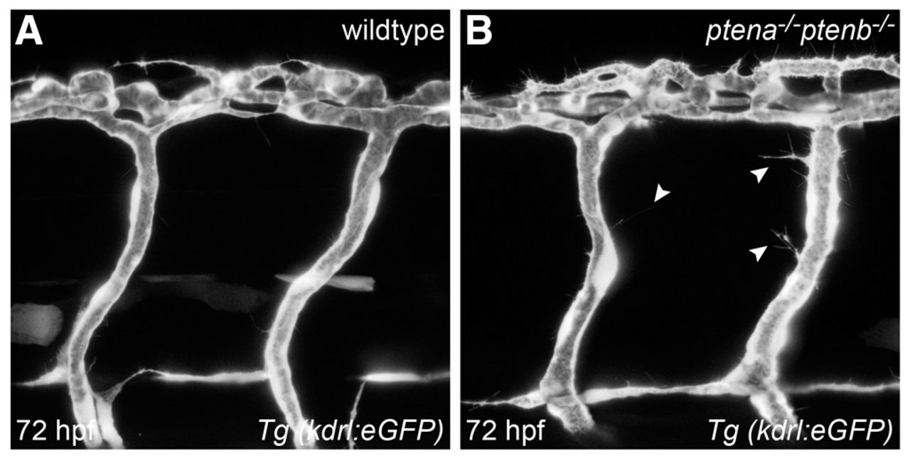
Loss of Ptena and Ptenb leads to excessive filopodia formation in endothelial cells at 72 hpf. Endothelial cells in living wild-type (A) and ptena-/-ptenb-/- mutant (B) embryos were visualized using Tg(kdrl:eGFP) and confocal imaging was performed at 70–72 hpf. Intersegmental vessels along the trunk in ptena-/-ptenb-/- mutants (4/4) show excessive filopodia formation (arrowheads), whereas no filopodia were observed in wild-type (0/4) embryos. Anterior to the left, 40× + 1.5 zoom, 0.5 μm step size.
Figure Data
Acknowledgments
This image is the copyrighted work of the attributed author or publisher, and
ZFIN has permission only to display this image to its users.
Additional permissions should be obtained from the applicable author or publisher of the image.
Full text @ Dis. Model. Mech.