
Fig. S2
- ID
- ZDB-IMAGE-131106-9
- Publication
- Hess et al., 2012 - Intravital Imaging of Thymopoiesis Reveals Dynamic Lympho-Epithelial Interactions
- All Figures
- Figures for Hess et al., 2012
|
Fig. S2
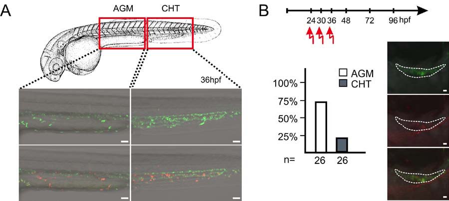
Time and place of origin of thymus-settling precursors
(A) Green fluorescent hematopoietic cells of fish transgenic for ikaros:tdEosFP were photoconverted in the indicated regions of the embryo (AGM, aorta-gonad-mesonephros; CHT, caudal hematopoietic tissue) at various time points (here shown for 36hpf); n=26 embryos for each time-point and region. Scale bars, 50μm.
(B) Colonization of the thymus by hematopoietic cells residing in the AGM or CHT regions photoconverted at 24, 30, or 36 hpf (time-line at top). No fluorescent cells are detectable after photoconversion at 24 and 30 hpf. The proportion of embryos with colonized thymi after photoconversion of the AGM or CHT at 36 hpf. Images of green, red and combined channels of a representative embryo after photoconversion in the AGM region are shown on the right. Scale bars, 10μm.
Reprinted from Immunity, 36(2), Hess, I., and Boehm, T., Intravital Imaging of Thymopoiesis Reveals Dynamic Lympho-Epithelial Interactions, 298-309, Copyright (2012) with permission from Elsevier. Full text @ Immunity