
Fig. S5
- ID
- ZDB-IMAGE-131105-46
- Publication
- Andersson et al., 2012 - Adenosine Signaling Promotes Regeneration of Pancreatic beta Cells In Vivo
- All Figures
- Figures for Andersson et al., 2012
|
Fig. S5
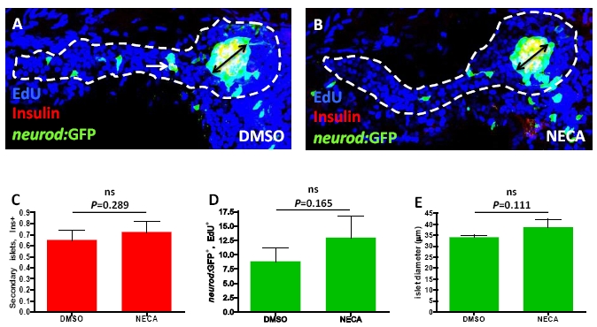
NECA treatment does not significantly affect pan-endocrine proliferation/morphology features during normal development, related to Figure 4.
(A-E) The transgenic line Tg(neurod:GFP) was used as a pan-endocrine marker in combination with Insulin staining and EdU incorporation between 4-6 dpf, to assess endocrine features at 6 dpf. (A) Confocal projection of a larva treated with DMSO from 4-6 dpf. Note that a secondary islet has formed (white arrow), and the diameter of the principal islet is marked (black arrow). (B) Confocal projection of a larva treated with NECA from 4-6 dpf. (C) No significant difference was found in the number of secondary islets when comparing DMSO- and NECA-treated larvae. (D) Quantification of the number of Tg(neurod:GFP)-expressing cells in the principal islet that had incorporated EdU revealed no significant difference between DMSO- and NECA-treated larvae. (E) The diameter of the principal islet did not significantly differ between DMSO- and NECA-treated larvae. n = 13 larvae for DMSO-treated, and n = 12 larvae for NECAtreated. Error bars represent SEM.
Reprinted from Cell Metabolism, 15(6), Andersson, O., Adams, B.A., Yoo, D., Ellis, G.C., Gut, P., Anderson, R.M., German, M.S., and Stainier, D.Y., Adenosine Signaling Promotes Regeneration of Pancreatic beta Cells In Vivo, 885-894, Copyright (2012) with permission from Elsevier. Full text @ Cell Metab.