
|
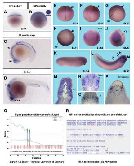
Fig. S1
lypd6 expression during zebrafish and Xenopus embryonic development and bioinformatic analyses of Lypd6 protein sequence.
related to Figure 1.
(A-D) Zebrafish lypd6 expression pattern detected by whole mount in situ hybridization (WMISH) at the indicated stages. ov = otic vesicle, opv = optic vesicle, hpf = hours post fertilization.
(E-P) Xenopus lypd6 expression pattern during embryogenesis. bpl = blastoporus lip, bp = blastoporus, npb = neural plate border, anp = anterior neural plate, ncc = neural crest cells, ov = otic vesicle. E, F, K and L lateral views, G and H vegetal views, I and J anterior views. M horizontal section, N-P transversal sections of embryos at stage 33 at the positions shown by dotted lines in L.
(Q) Signal peptide prediction in zebrafish Lypd6 using Signal P 4.0 Server (University of Denmark). Cleavage site predicted between position 22 and 23: IKA-VQ.
(R) GPI-anchor modification site prediction in zebrafish Lypd6 using big-PI Predictor (I.M.P. Bioinformatics). GPI-anchor attachment site (Omega site) predicted at position 147: N.
Reprinted from Developmental Cell, 26(4), Özhan, G., Sezgin, E., Wehner, D., Pfister, A.S., Kühl, S.J., Kagermeier-Schenk, B., Kühl, M., Schwille, P., and Weidinger, G., Lypd6 Enhances Wnt/beta-Catenin Signaling by Promoting Lrp6 Phosphorylation in Raft Plasma Membrane Domains, 331-345, Copyright (2013) with permission from Elsevier. Full text @ Dev. Cell