
|
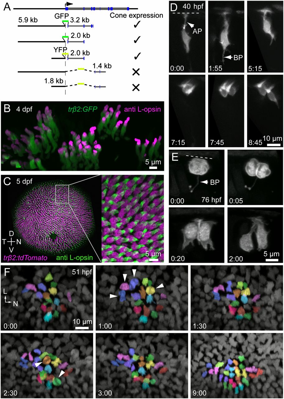
Fig. 1
L cones are derived from symmetric precursor divisions. (A) Intron-1 of zebrafish trβ2 is essential for expression in cones. (B) Transient expression of FP driven by the trβ2 promoter is restricted to cones immunopositive for L-opsin. L-opsin signal is restricted to the outer segment, where GFP labeling is dim because there is little cytoplasmic space in this compartment (also see Fig. S1). L-opsin signal in GFP-expressing cells were isolated by digitally removing the opsin signal outside the GFP masks. (C) Only L cones are labeled in the stable transgenic line, Tg(trβ2:tdTomato). D, dorsal; N, nasal; T, temporal; V, ventral. (D) In vivo multiphoton time-lapse imaging reveals division of a trβ2:MYFP-labeled cell that produced two cones. (Time, hours:minutes.) AP, apical process; BP, basal process. The dotted line indicates the location of the outer limiting membrane (also in E). Cells were labeled by transient expression of trβ2:MYFP. (E) Imaging at more frequent intervals shows that the dividing cell has a short process that bifurcates at its terminal ending (BP). Cells were labeled by transient expression of trβ2:MYFP-2A-histone2AYFP. (F) Snapshots from a time-lapse sequence demonstrating the generation of L cones in a Tg(trβ2:tdTomato) fish. See Movie S1 for the full sequence. Postmitotic cones already present within the patch were not tracked. Each color represents a pair of L cones produced from a terminal division during the imaging period. Arrowheads indicate mitotic figures. L, lateral; N, nasal.