
Fig. S2
- ID
- ZDB-IMAGE-131029-10
- Genes
- Publication
- Neugebauer et al., 2013 - Differential roles for 3-OSTs in the regulation of cilia length and motility
- All Figures
- Figures for Neugebauer et al., 2013
|
Fig. S2
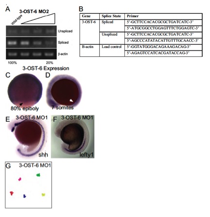
Splice-blocking efficacy, primers used to assay splicing, tracks of fluorescent beads injected into the KV at 6 SS, 3-OST-6 RNA expression, midline marker expression. (A) Analysis of 3-OST-6 MO2 splice-blocking efficacy. Increasing amounts of MO injected resulted in an 80±20% decrease of spliced mRNA. Analysis was performed as described in Fig. S1. (B) Table containing primers used for detecting spliced versus unspliced transcript when embryos were injected with translational splice blocking morpholinos. (C,D) Whole mount in situ analysis of 3-OST-6 mRNA, which was expressed ubiquitously throughout the embryo during epiboly (C) and early somitogenesis (D). Black arrow indicates staining in DFCs, white arrow indicates the position of KV. (E) Whole-mount in situ analysis of shh expression, which was normal in 3-OST-6 morphants (n=109). (F) Whole-mount in situ analysis of lefty1 expression in the midline, was normal in 3-OST6 morphants (n=49). (G) Bead tracks of fluorescent beads injected into 3-OST-6 MO1 embryos showing a lack of bead movement, indicative of nonmotile cilia.