
Fig. 2
- ID
- ZDB-IMAGE-130911-2
- Publication
- Miyake et al., 2013 - Fgf22 regulated by Fgf3/Fgf8 signaling is required for zebrafish midbrain development
- All Figures
- Figures for Miyake et al., 2013
|
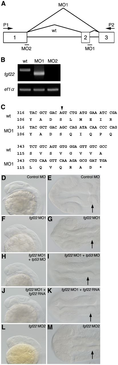
Fig. 2 Inhibition of fgf22 functions in zebrafish embryos.
(A) The coding region of fgf22 is divided by two introns. Open boxes and black lines indicate exons and introns, respectively. MO indicates the target position of fgf22 MO. (B) fgf22 cDNA was amplified from wild-type or fgf22 MO-injected embryonic cDNA by RT-PCR using P1 and P2 primers, the positions of which are indicated by arrows (A). ef1α cDNA was also amplified as a control. (C) The nucleotide sequences of fgf22 cDNAs described above were determined. Numbers for the nucleotide sequence of the coding region and the amino acid sequence are shown. Arrowheads indicate splice-sites between exons one and two. (D–M) Lateral views (D,F,H,J,L) and dorsal views (E,G,I,K,M) of control MO-injected (D,E), fgf22 MO1-injected (F,G), fgf22 MO1- and tp53 MO-injected (H,I), fgf22 MO1- and fgf22 RNA-injected (J,K), and fgf22 MO2-injected (L,M) embryos at 24 hpf are shown.