
Fig. 5
- ID
- ZDB-IMAGE-130910-17
- Publication
- Kim et al., 2013 - Multi-organ Abnormalities and mTORC1 Activation in Zebrafish Model of Multiple Acyl-CoA Dehydrogenase Deficiency
- All Figures
- Figures for Kim et al., 2013
|
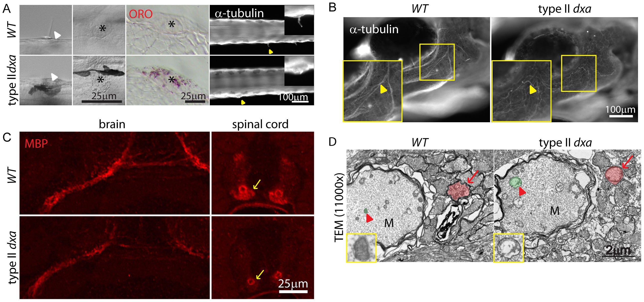
Fig. 5
Facial axons, mechanosensory hair cell and myelination defects in type II dxavu463 mutant zebrafish.
(A) DIC imaging of live cilia (first column) and neuromast cells (second column, asterisks), ORO stained lipids in neuromast cells (third column, asterisks). Acetylated-tubulin marks cilia (yellow arrowheads). Magnified views of cilia are shown on the upper right corner. (B) Whole mount immunofluorescence staining of acetylated-tubulin in WT (left) and dxa mutants (right) at 8 dpf. Yellow arrowheads indicate facial axons. Rectangle region is magnified in lower left corner. (C) anti-MBP staining in the brain (left) and spinal cord (right) in WT (top) and dxa mutant zebrafish (bottom) at 8 dpf. Arrows indicate myelinated axons in the spinal cord, all signal is reduced in dxa mutant zebrafish. (D) TEM (11,000×) image of spinal cord as indicted by arrows in C. Normal (WT) and swollen (dxa mutant) mitochondria are pseudocolored green, indicated by large red arrowhead). Red arrows indicate less condensed myelination layer in a dxa axon. Further magnified views of mitochondria are on the lower left corner. M, Mauthner axon track. Scale bars are as indicated in each panel.