
Fig. S1
- ID
- ZDB-IMAGE-130907-9
- Publication
- Alunni et al., 2013 - Notch3 signaling gates cell cycle entry and limits neural stem cell amplification in the adult pallium
- All Figures
- Figures for Alunni et al., 2013
|
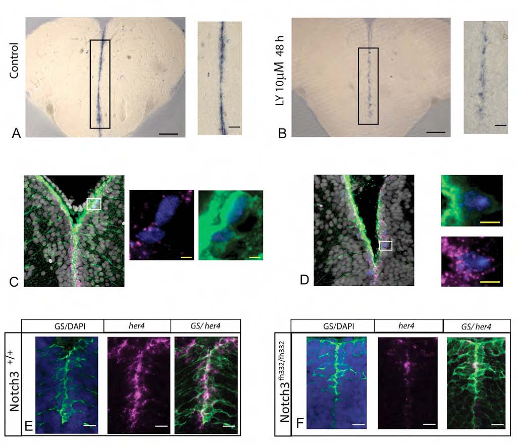
Fig. S1
her4.1 expression depends on Notch signaling in adult pallial RG, and is a Notch3 target in RG of the juvenile pallium.
(A,B) Expression of her4.1 revealed by in situ hybridization (blue) in a control brain or following 48 hours of LY treatment. Notch blockade reduces her4.1 expression along the ventricular zone. Boxed areas are enlarged on the right of each panel. (C,D) her4.1 expression revealed by fluorescent in situ hybridization (magenta) on telencephalic cross-sections of a gfap:gfp brain, together with a fluorescent immunostaining for MCM5 (blue). her4.1 transcripts mostly characterize non-dividing RG cells, and only in rare cases dividing RG cells. (D) Boxed areas are enlarged on the right of each panel. (C) Two examples of her4.1-negative or weakly dividing RG. (D) One example of a her4.1-positive dividing RG. (E,F) Expression of her4.1 revealed by fluorescent in situ hybridization (magenta) together with immunostaining for the RG marker GS (green) (counterstained for DAPI, blue) at 5 dpf in notch3 fh332fh332 homozygotes (F) and wild-type siblings (E). her4.1 expression is strongly reduced in notch3 fh332fh332 mutants compared with wild-type siblings. Scale bars: 100 μm (black); 10 μm (white); 4 μm (yellow).