Fig. S3
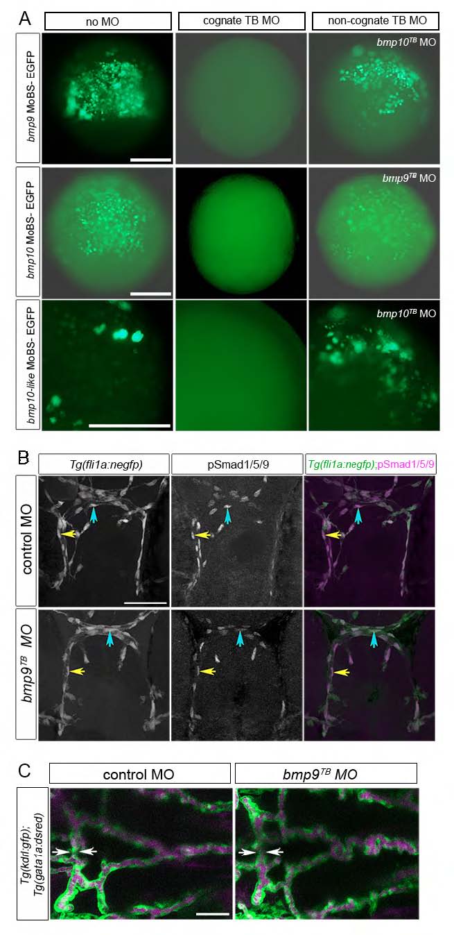
bmp9 knockdown does not phenocopy zebrafish alk1-/- mutants. (A) Translation blocking (TB) morpholino validations. Wild-type embryos injected at the one-cell stage with 50 pg CMV-driven EGFP constructs modified with morpholino-binding sites (MoBS) inserted upstream of the ATG, with or without cognate or non-cognate morpholinos. bmp9TB MO, 7 ng; bmp10TB MO, 20 ng; bmp10-like MO, 3 ng. Embryos were observed at ~6 hpf for EGFP fluorescence. Scale bar: 500 μm (top two rows) or 250 μm (bottom row). (B) pSmad1/5/9 expression (middle column) in endothelial cells (nuclei marked by fli1a:negfp transgene, left column) in 36 hpf embryos injected with 7 ng control or bmp9TB morpholino. Higher amounts of this morpholino were overtly toxic. In merge (right column), EGFP-expressing endothelial cell nuclei are green, pSmad1/5/9 immunofluorescence is magenta. Yellow and blue arrows indicate endothelial cells in the CaDI and BCA, respectively. 2D confocal projections of 50 μm frontal sections, dorsal upwards. Scale bar: 50 μm. See Table S1 for fluorescence quantitation. (C) Cranial vasculature in 48 hpf Tg(kdrl:gfp);Tg(gata1a:dsRed) embryos injected with 7 ng control or bmp9TB morpholino. Arrows highlight width of BCA. Endothelial cells are green, red blood cells are magenta. 2D confocal projections, dorsal views, anterior leftwards. Scale bar: 50 μm. n=207 over five experiments.

