
Fig. S3
- ID
- ZDB-IMAGE-130907-11
- Publication
- Alunni et al., 2013 - Notch3 signaling gates cell cycle entry and limits neural stem cell amplification in the adult pallium
- All Figures
- Figures for Alunni et al., 2013
|
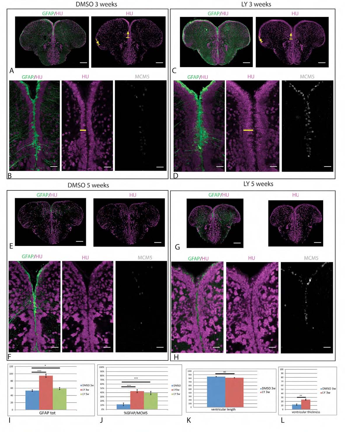
Fig. S3
LY treatments of 3 and 5 weeks highlight a regulation of RG amplification but the maintenance of a NSC zone. (A-H) Cross-sections of the adult pallial ventricular zone in gfap:gfp transgenic brains processed in triple immunocytochemistry for the detection of GFP (RG, green), HuC/D (magenta) and MCM5 (gray). Adult fish were continuously treated with LY (or DMSO for controls) for 3 weeks (A-D) or 5 weeks (E-H). Low magnification panels (A,C) were used to measure the pallial ventricular surface (between arrows, see K). The horizontal yellow bar in B,D indicates the width of the subventricular neuronal domain. Scale bar: 10 μm. Confocal projection images from four optical planes. (I,J) Quantification of the total number of RG cells per section (I) and the proportion of RG in proliferation (J) following control (blue), 3-week (red) or 5-week (green) treatments. (K,L) Quantification of the pallial ventricular length (K) and width of the subventricular neuronal population (L) following control (blue) and 3-week (red) treatments. *P<0.05; **P<0.001; ***P<0.0001 (n=3 brains for each condition).