
Fig. S1
- ID
- ZDB-IMAGE-130906-33
- Publication
- Sorrell et al., 2013 - Tcf7l1 proteins cell autonomously restrict cardiomyocyte and promote endothelial specification in zebrafish
- All Figures
- Figures for Sorrell et al., 2013
|
Fig. S1
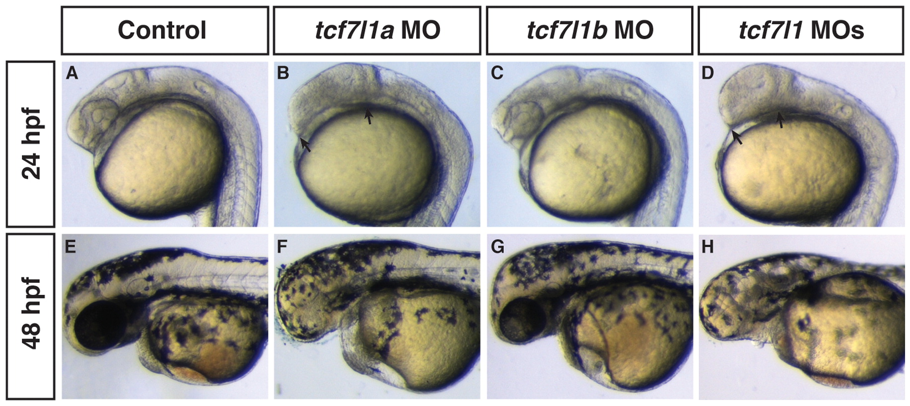
Efficacy of Tcf7l1 MOs demonstrating a functional interaction of Tcf7l1a and Tcf7l1b in promoting the anterior neural tissue. (A,E) Control embryos. (B,F) Tcf7l1a deficient embryos. (C,G) Tcf7l1b deficient embryos. (D,H) Tcf7l1 deficient embryos. We observed anterior neural patterning defects and functional interactions between Tcf7l1a and Tcf7l1b exactly as has been reported previously (Dorsky et al., 2003). Arrows in B and D indicate the length between the midbrain-hindbrain boundary and the anterior head, which is significantly shorter in the Tcf7l1 deficient embryos compared to the Tcf7l1a deficient embryos.
Reprinted from Developmental Biology, 380(2), Sorrell, M.R., Dohn, T.E., D'Aniello, E., and Waxman, J.S., Tcf7l1 proteins cell autonomously restrict cardiomyocyte and promote endothelial specification in zebrafish, 199-210, Copyright (2013) with permission from Elsevier. Full text @ Dev. Biol.