
|
Fig. 4
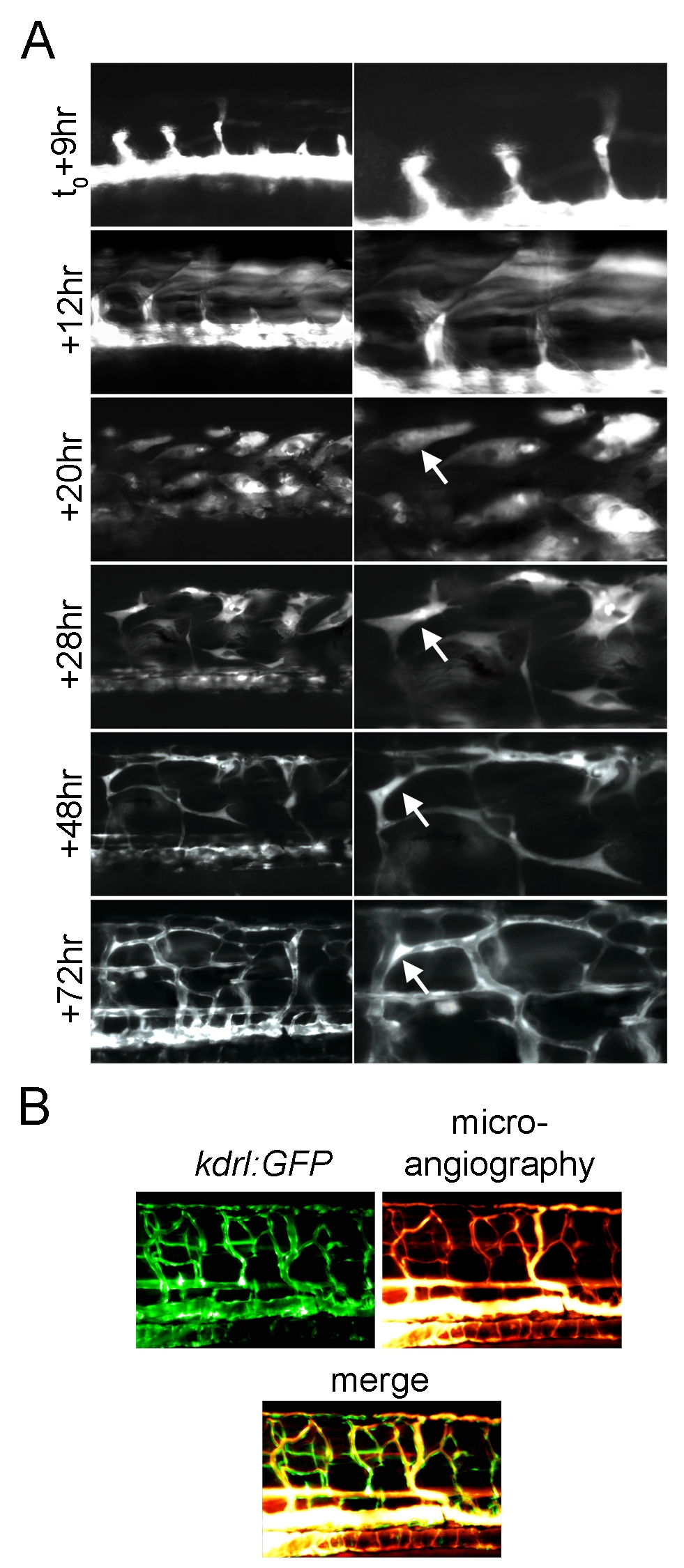
Fast skeletal muscle converts to functional endothelial cells following Etv2 expression.
(A) Extended time lapse imaging of the trunk of a kdrl:GFP/hsp70l:etv2 embryo showing ectopic GFP expressing cells change morphology from fiber-like (+12 h) to spindle-like (+20 h, +28 h) to form a network of thin cells (+48 h) and finally appear to form lumenized vessels (+72 h). The two panels shown at each time point are two magnifications of the same image. (B) Microangiography demonstrates the newly formed vessels are functional. Rhodamine dextran dye was injected into the circulation of a 4 dpf (+72 h post–heat shock) kdrl:GFP/hsp70l:etv2 embryo. Rhodamine labels within all of the newly formed vessels and no vascular leakage are observed.