
|
Fig. 2
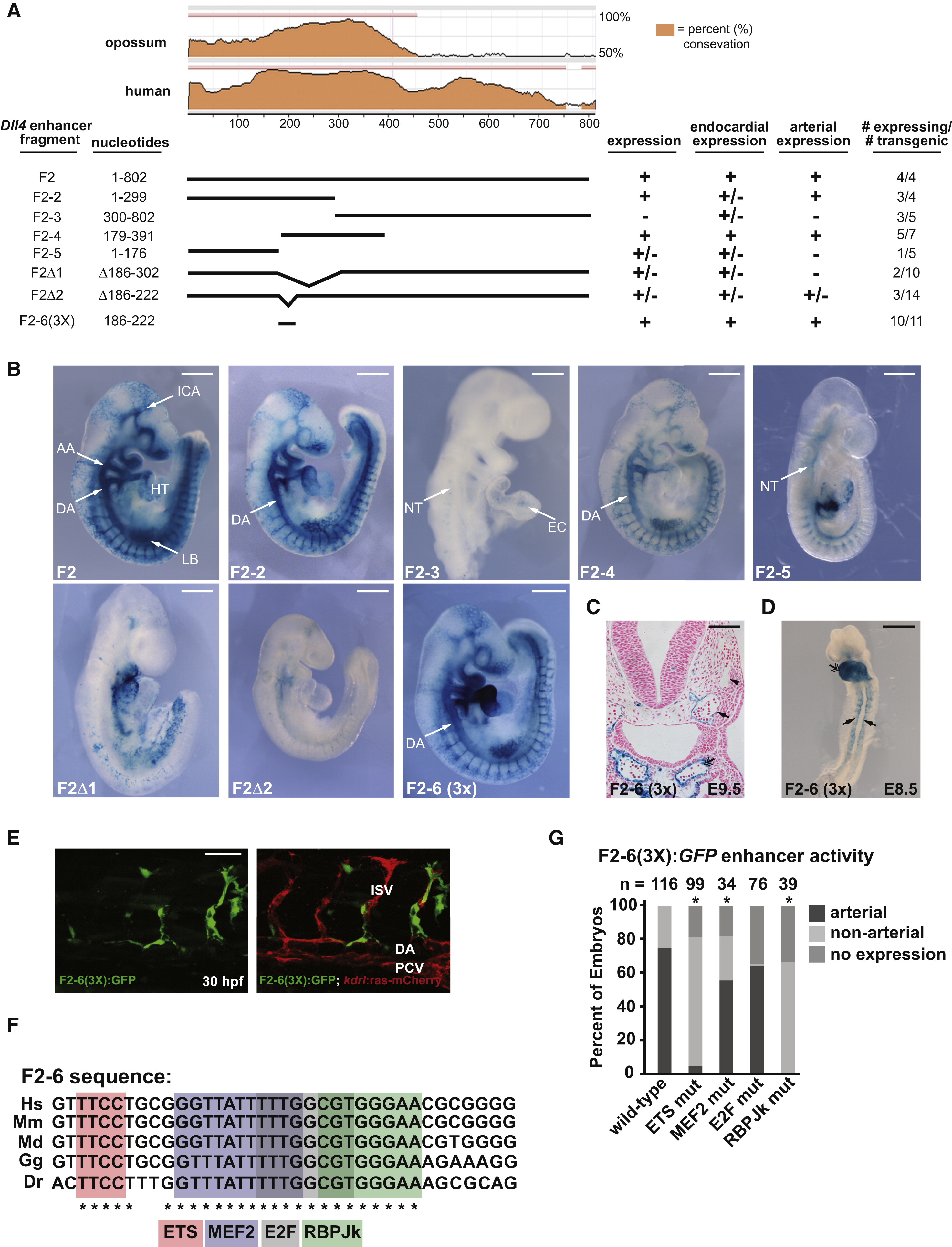
Isolation of a Minimal Dll4 Enhancer Element that Drives Arterial-Specific Expression (A) Sequence conservation of the F2 enhancer and deletion constructs used in transgenic analyses. Endocardial and arterial expression is indicated. (B) Representative transgenic embryos from (A) at E9.5. F2, F2-2, and F2-4 directed strong arterial-specific expression. F2-3 directed weak endocardial (EC) expression. Deletion of a highly conserved 100 bp and 36 bp region (F2Δ1 and F2Δ2, respectively) abrogated arterial activity of the F2 enhancer. The 36 bp region, F2-6, was sufficient, when arrayed in triplicate, F2-6(3X), to direct arterial expression. DA, dorsal aorta; AA, aortic arch; LB, limb bud; NT, neural tube; ICA, internal cerebral artery; HT, heart. Scale bar represents 500 μm. (C) Transverse section of X-gal stained F2-6(3X)-lacZ embryo at E9.5. DA (arrow); cardinal vein (caret); EC (double arrow). Scale bar represents 100 μm. (D) Whole-mount image of F2-6(3X)-lacZ embryo at E8.5. Scale bar represents 500 μm. (E) Mosaic expression of F2-6(3x):GFP in the DA and intersomitic vessels (ISVs) of a transient transgenic 30 hpf zebrafish embryo. PCV, posterior cardinal vein. Scale bar represents 50 μm. (F) Sequence comparison of F2-6 in human (Hs), mouse (Mm), opossum (Md), chicken (Gg), and zebrafish (Dr). Cis elements are indicated. (G) Each cis element was mutated in F2-6(3X):GFP and transient transgenics were assessed for arterial expression at 24 hpf. The percentage of embryos with expression in arteries (i.e., DA and/or ISV), expression elsewhere, and no expression, are indicated. Asterisk indicates a significant difference in arterial expression compared to wild-type (χ2 test). See also Figure S2.
Reprinted from Developmental Cell, 26(1), Wythe, J.D., Dang, L.T., Devine, W.P., Boudreau, E., Artap, S.T., He, D., Schachterle, W., Stainier, D.Y., Oettgen, P., Black, B.L., Bruneau, B.G., and Fish, J.E., ETS Factors Regulate Vegf-Dependent Arterial Specification, 45-58, Copyright (2013) with permission from Elsevier. Full text @ Dev. Cell